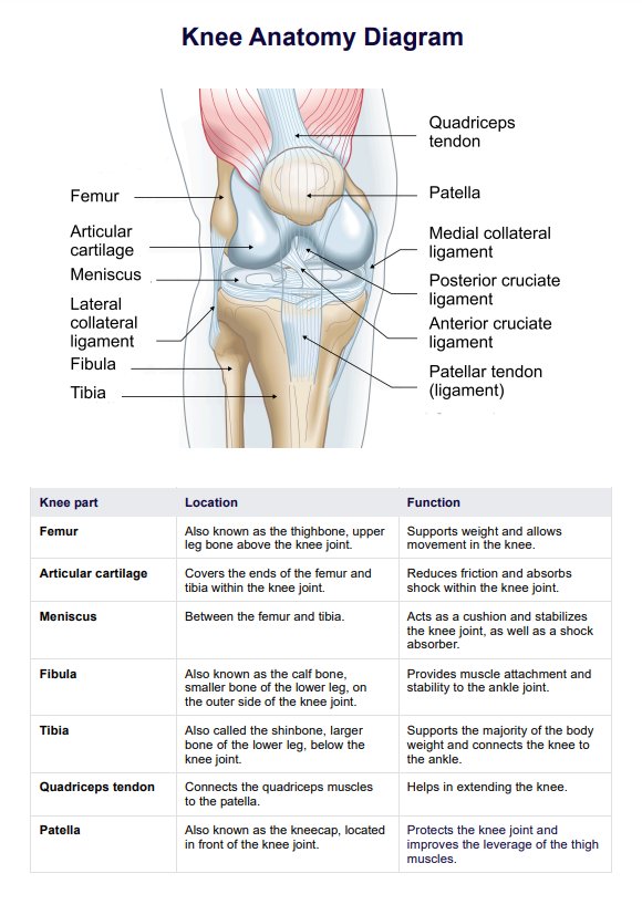
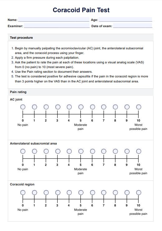
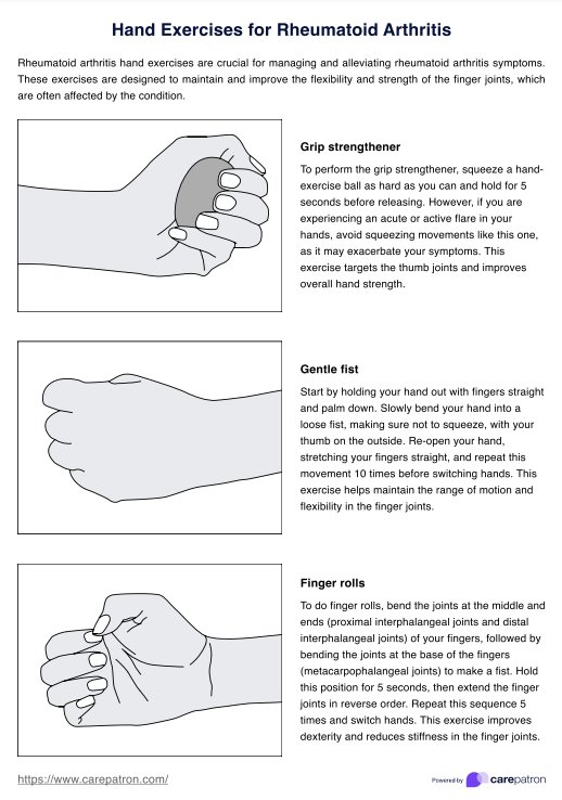
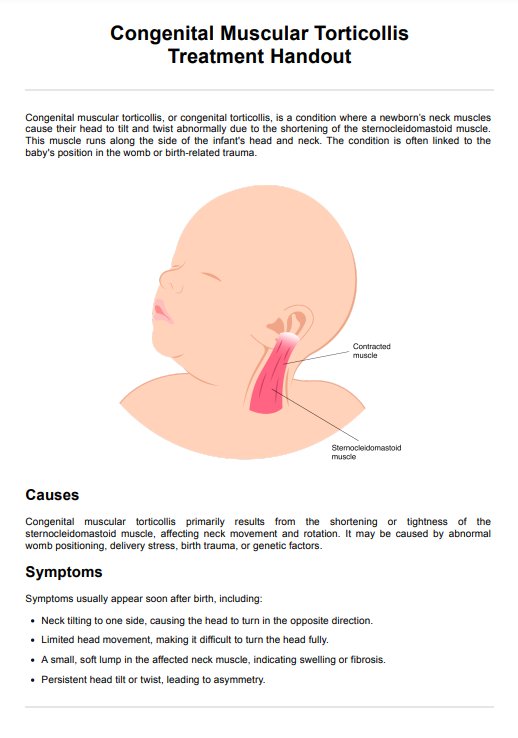
Gallbladder Physical Exam
Learn about the physical exam for the gallbladder, including techniques and examples. Download Carepatron's free gallbladder physical exam PDF guide for reference.

Fact Checked by RJ Gumban.


What is a Gallbladder Physical Exam?
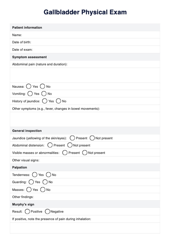
A Gallbladder Physical Exam is a clinical assessment focused on the abdomen, particularly the right upper quadrant, to identify signs of gallbladder disease. The patient is typically positioned supine, and the healthcare provider begins with a visual inspection for signs such as jaundice, abdominal distension, or any visible masses.
Palpation of the abdomen is crucial for assessing tenderness, especially in the right upper quadrant, which can indicate conditions like gallstones or cholecystitis (inflammation of the gallbladder). Another key component of the exam is Murphy's sign, where the provider applies pressure to the gallbladder area while the patient takes a deep breath; a sharp increase in pain suggests gallbladder inflammation. The provider will also inquire about symptoms such as abdominal pain, nausea, vomiting, and jaundice, which are common indicators of gallbladder issues.
Additional signs, such as Courvoisier's sign (a palpable gallbladder due to bile duct obstruction) and signs of guarding or rebound tenderness, may also be used for assessment. Overall, this physical exam is essential for diagnosing gallbladder diseases and guiding further evaluation and treatment.
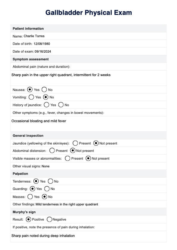
Gallbladder Physical Exam Template
Gallbladder Physical Exam Example
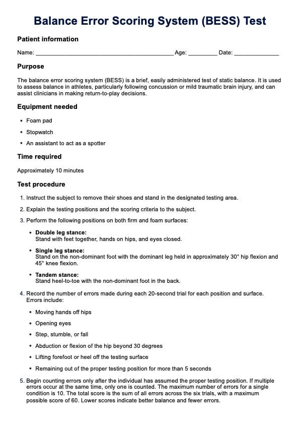
How to conduct a Gallbladder Physical Exam
Conducting a physical exam for the gallbladder involves a systematic approach to assessing signs and symptoms associated with gallbladder disease. Here are the key steps to guide you through the process:
Step 1: Assessment of symptoms
Inquire about the patient's symptoms, including the nature and duration of abdominal pain, nausea, vomiting, and history of jaundice.
Step 2: General inspection
Begin with a general inspection of the abdomen. Look for any signs of jaundice, abdominal distension, or visible masses. Note any visual abnormalities that may provide initial clues about the patient's abdominal health.
Step 3: Palpation
Palpate the abdomen, particularly the right upper quadrant, to assess for tenderness, guarding, or masses
Step 4: Murphy's sign
Press on the gallbladder area while the patient takes a deep breath. A positive Murphy's sign indicates tenderness and may suggest acute cholecystitis if the patient experiences increased pain during inhalation.
Step 5: Document findings
Record any significant findings using the Gallbladder Physical Exam template. Our template includes sections for documenting the patient's symptoms, general inspection, palpation results, and any additional notes or observations. This documentation will help you keep track of your findings and assist in making a diagnosis.
Types of gallbladder disease
Gallbladder disease encompasses a range of conditions that affect the proper functioning of this vital organ. Understanding these different types is crucial for identifying symptoms and seeking appropriate medical attention. Let's explore the primary types of gallbladder diseases:
Biliary colic
Biliary colic is characterized by intense pain in the upper abdomen, typically caused by the temporary obstruction of the bile ducts. This occurs when gallstones, small solid particles that can form in the gallbladder, obstruct the normal flow of bile. The resulting pain can be sharp and cramp-like, often triggered by the consumption of fatty foods.
Acute cholecystitis
Acute cholecystitis is an inflammation of the gallbladder, often stemming from gallstones' blockage of the cystic duct. The condition can cause persistent upper abdominal pain, tenderness, and swelling. Physical findings associated with acute cholecystitis may include a positive Murphy's sign—a sharp increase in pain upon deep palpation of the gallbladder area.
Gallstones
While not a disease per se, the formation of gallstones can lead to various gallbladder-related complications. These solid particles, primarily composed of cholesterol or bilirubin, can range in size and obstruct the normal flow of bile. Abdominal pain in the upper part is a common symptom, often radiating to the back or right shoulder.
Gallstone disease symptoms
Gallstone disease often presents with a spectrum of symptoms, and understanding these signs is vital for early detection and intervention. Here are some critical symptoms associated with gallstone disease:
Upper abdominal pain
Abdominal pain in the upper area is a hallmark symptom of gallstone disease. Patients frequently describe a persistent, gnawing discomfort in the upper right quadrant, often radiating toward the back or right shoulder. During a physical examination, healthcare professionals may inquire about the nature, intensity, and duration of this pain to aid in diagnosis.
Nausea and vomiting
Nausea and vomiting are common accompanying symptoms. These symptoms may arise due to the disruption of normal bile flow caused by gallstones. A thorough physical examination includes assessing the patient's history of nausea and vomiting with pain in the upper abdominal episodes.
Jaundice
Jaundice is a yellowing of the skin and eyes resulting from the accumulation of bilirubin. While gallstones primarily impact the gallbladder, common bile duct stones can obstruct bile flow and lead to jaundice. A careful examination may involve inspecting the skin and sclera for signs of yellow discoloration.
How to diagnose gallstone disease
Diagnosing gallstone disease involves a combination of clinical evaluation and diagnostic tests to confirm the presence of gallstones and assess their impact on the gallbladder and surrounding structures. Here are some ways how to diagnose gallstone disease, according to the National Institute of Diabetes and Digestive and Kidney Diseases (2017):
Laboratory tests
Laboratory tests are crucial in diagnosing gallstone disease by providing insights into liver function and identifying markers associated with gallbladder issues. Common tests include:
- Liver function tests (LFTs): Assessing liver enzymes such as alanine aminotransferase (ALT) and alkaline phosphatase (ALP) can help identify any liver dysfunction associated with gallstone-related complications.
- Bilirubin levels tests: Elevated bilirubin levels may indicate common bile duct obstruction, a complication of gallstones. Total and direct bilirubin levels are assessed to gauge the severity of the obstruction.
- Amylase and lipase levels tests: While primarily associated with pancreatitis, elevated amylase and lipase levels may suggest gallstone-induced inflammation affecting the pancreas.
Imaging studies
Accurate diagnosis also often uses advanced imaging techniques to visualize the gallbladder and associated structures. The following imaging studies are commonly employed:
- Ultrasound: Ultrasonography is a widely used imaging technique for visualizing the gallbladder and detecting the presence of gallstones. It helps assess the size, number, and location of stones and any associated inflammation.
- CT scan: In cases where ultrasound results are inconclusive or additional information is needed, a computed tomography (CT) scan may be recommended. CT scans provide detailed cross-sectional images of the abdomen, aiding in diagnosing complications like inflammation or infection.
- Endoscopic retrograde cholangiopancreatography (ERCP): ERCP is an invasive procedure that combines endoscopy and fluoroscopy to visualize the bile ducts. It is instrumental in identifying and removing gallstones lodged in the common bile duct, offering diagnostic and therapeutic benefits.
- Cholescintigraphy (HIDA scan): A HIDA (hepatobiliary) scan involves injecting a radioactive tracer into the bloodstream, which is then taken up by the liver and excreted into the bile. This helps assess the gallbladder's function, identifying issues such as gallstones or obstruction.
Reference
National Institute of Diabetes and Digestive and Kidney Diseases. (2017, November). Diagnosis of gallstones | NIDDK. https://www.niddk.nih.gov/health-information/digestive-diseases/gallstones/diagnosis
Commonly asked questions
A physical exam of the gallbladder involves a healthcare professional assessing the abdomen, specifically the right upper quadrant, through palpation and observation to identify signs of tenderness, inflammation, or abnormalities associated with gallbladder issues.
Collin's sign is a clinical finding observed during a physical exam of the gallbladder. It refers to tenderness in the right subcostal area upon inspiration, often indicative of gallbladder inflammation or cholecystitis.
Murphy's sign on a sonograph involves observing for a pause in inspiration due to pain when the ultrasound transducer is pressed over the gallbladder area. This can indicate gallbladder inflammation or obstruction, providing valuable diagnostic information.
Related Templates

Popular Templates