Chest Radiograph
Explore our detailed guide on Chest Radiography, offering insights into effective diagnostic techniques for various thoracic conditions. Download the free PDF now.

Fact Checked by Ericka Pingol.


What is a Chest Radiograph?
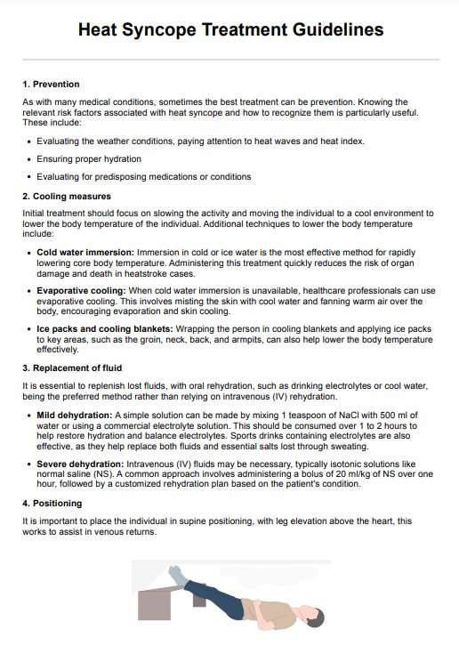
A Chest Radiograph commonly known as a chest X-ray, is a diagnostic imaging test that uses a small amount of ionizing radiation to produce images of the structures inside the chest, including the heart, lungs, airways, blood vessels, and bones of the spine and chest area. This test is one of the most common imaging tests performed due to its effectiveness in diagnosing, monitoring, and treating various conditions affecting the chest.
Purpose
The primary purpose of Chest Radiography is to evaluate the chest organs and structure, aiding in diagnosing conditions affecting the lungs, heart, and chest wall. It is particularly useful in detecting pneumonia, heart failure, emphysema, lung cancer, and other medical conditions.
Chest X-rays also help assess symptoms such as cough, shortness of breath, or chest pain. These can identify cardiac abnormalities, including an enlarged heart, which may indicate congestive heart failure or other heart diseases.
When to perform a Chest Radiograph
A Chest Radiograph is typically performed when a patient presents symptoms of a chest infection, persistent cough, chest pain, injury, or difficulty breathing. It is also used as a routine diagnostic tool for pre-surgical preparation or to monitor the progression of diagnosed diseases such as tuberculosis or heart failure.
Additionally, healthcare providers may order a chest X-ray to check the placement of devices like pacemakers or catheters. For detailed protocols on assessing chest pain, you can download our Chest Pain Workup template, designed to assist healthcare providers in systematic evaluation.
How to perform a Chest Radiograph
Performing a Chest Radiograph involves positioning the patient either standing or sitting in front of an X-ray machine. The patient is usually asked to hold their breath while the X-ray image is taken to avoid blurring. Two views, a frontal view, and a lateral view, are typically taken for a complete assessment. The procedure is quick, usually lasting only a few minutes, and does not require any special preparation. The radiologist then reviews the X-ray image and provides a detailed report on the findings.
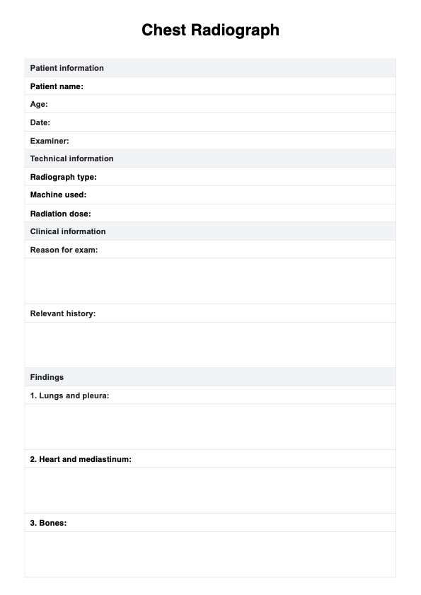
Chest Radiograph Template
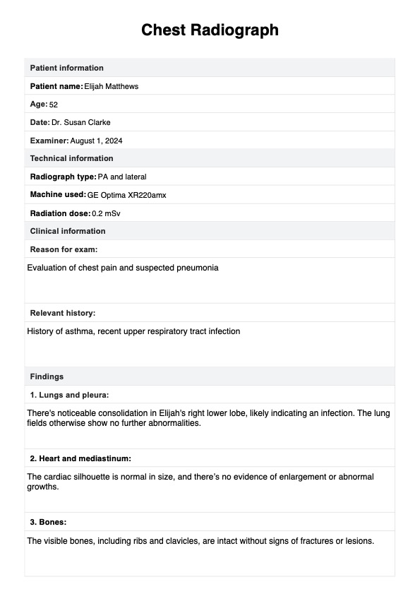
Chest Radiograph Example
Types of Chest Radiograph projections
Chest Radiograph projections refer to the different angles and positions in which X-ray images of the chest are captured to provide the best view of the anatomical structures. The choice of projection depends on the clinical question, the patient's condition, and their ability to assume different positions. Here are the main types of Chest Radiograph projections:
- Posteroanterior (PA) projection: This is the standard and most commonly used chest X-ray view. The patient stands facing an X-ray plate, with the X-ray beam passing from back to front. This projection gives a clear view of the heart, lungs, and major vessels, minimizing the magnification of the heart.
- Lateral projection: Used in conjunction with the PA view, the patient stands with arms raised and the side of their chest against the X-ray plate. This view helps to localize abnormalities seen on the PA projection by providing a side perspective.
- Anteroposterior (AP) projection: This projection is often used when patients cannot stand, such as those in intensive care or bedridden. The X-ray beam passes from front to back while the patient is lying down or sitting. Compared to the PA projection, this projection can magnify the view of the heart and other structures.
Preparations for a Chest Radiograph
Preparing for a Chest Radiograph is generally straightforward, involving minimal steps to ensure accurate and clear imaging results. Here are the key preparations:
- Clothing: Patients are usually asked to remove all clothing and jewelry from the waist up to prevent any obstructions in the images. They are typically provided with a hospital gown to wear during the procedure.
- Positioning: Proper positioning is crucial for a successful chest X-ray. Patients may be asked to stand before the X-ray machine and hold their breath for a few seconds as the image is taken. This prevents blurring and ensures the lungs are well-expanded.
- Medical history: Patients should inform the radiologist or technician about any recent surgeries, the presence of implants, or other medical conditions. This information can help adjust the technique or evaluate the images more effectively.
- Pregnancy: Women should always inform the medical team if there is any possibility that they are pregnant, as precautions may be needed to minimize radiation exposure to the fetus.
Technical assessments of a Chest Radiograph
Technical assessments of chest radiographs are essential for ensuring the quality and diagnostic effectiveness of the images produced. Key criteria for evaluating these radiographs include penetration, inspiration, rotation, magnification, and angulation ((Bickle & Manickam, 2018; Mowery & Singh, 2024):
Penetration
This refers to the amount of radiation that passes through the patient's chest during the imaging process. Over-penetration can result in a dark, under-exposed image, while under-penetration can lead to a light, over-exposed image. This can affect the visibility and clarity of structures within the chest, making it difficult to identify any abnormalities.
Inspiration
Proper inspiration is crucial in obtaining a quality Chest Radiograph. Deep inspiration helps to expand the lungs and separate the ribs, allowing for better visualization of the lung fields and diaphragm. A patient who is unable to fully inspire may have a collapsed lung or underlying respiratory condition that should be noted by the radiologist.
Rotation
Rotation refers to the position of the patient's body in relation to the image receptor. Proper rotation is important as it ensures that all structures within the chest are aligned symmetrically, making it easier to identify any abnormalities. Improper rotation can result in distorted images with asymmetrical structures, leading to potential misinterpretation.
Magnification
Magnification occurs when there is an increase in size of structures on an image due to improper distance between the patient and image receptor. This can affect measurements and make it difficult to accurately identify any abnormalities.
Angulation
Angulation refers to the angle at which the x-ray beam is directed towards the patient. Proper angulation is important as it allows for better visualization of structures and reduces radiation exposure. Improper angulation can result in distorted images or missed abnormalities.
What can a Chest Radiograph detect and help diagnose?
A Chest Radiograph, commonly known as a chest X-ray, is a versatile diagnostic tool used in medicine to examine the structures inside the chest. This imaging technique is crucial for detecting various conditions and abnormalities within the chest cavity.
- Respiratory conditions: Chest Radiographs are instrumental in diagnosing respiratory diseases such as pneumonia, tuberculosis, and chronic obstructive pulmonary disease (COPD). They can reveal lung infections through visible infiltrates or fluid in the lungs and help monitor the progression of chronic lung conditions.
- Cardiovascular issues: Chest X-rays can identify cardiac abnormalities, including an enlarged heart, which may indicate heart failure or other heart diseases. They are also used to detect abnormalities in the size and shape of the heart and major blood vessels, providing essential clues about cardiovascular health.
- Bone and structural problems: Chest Radiographs can show abnormalities in the bones of the chest wall, spine, and ribs, such as fractures from a chest injury, providing crucial diagnostic information. They are particularly useful in assessing traumatic injuries following accidents.
- Other conditions: These imaging tests can detect certain types of tumors within the chest, the presence of fluid or air around the lungs, and diaphragmatic hernias. They are also valuable for checking the placement and condition of medical devices, like pacemakers, catheters, and other implants.
Commonly asked questions
A Chest Radiograph can provide valuable information about the heart, lungs, airways, bones of the chest and spine, and the diaphragm, helping to detect conditions such as pneumonia, heart failure, fractures, and tumors.
A Chest Radiograph is a quick procedure, typically taking only a few minutes to complete, as the actual exposure to radiation lasts only a fraction of a second.
The benefits of a Chest Radiograph include its speed and ease of use, the minimal radiation exposure compared to other imaging forms, and its effectiveness in diagnosing a wide range of chest and lung conditions, which can lead to timely and appropriate medical treatment.
Related Templates

Popular Templates