Cavovarus Foot Treatment Guidelines Handout

Discover the benefits of our Cavovarus Foot Treatment Guidelines Handout for healthcare professionals. Download now and enhance patient care with this comprehensive PDF.

By Emma Hainsworth on Dec 11, 2024.

tick

Fact Checked by Ericka Pingol.

Use Template
AI IconToolbarShare ui

What is a cavovarus foot?

A cavovarus foot is a structural deformity characterized by an abnormally high medial arch (pes cavus) combined with an inward tilt of the heel (hindfoot varus). This deformity disrupts the biomechanics of the foot, leading to uneven weight distribution and altered gait mechanics. The condition can vary in severity, ranging from mild flexibility to rigid deformities. Cavovarus feet often cause instability, pain, callus formation, and in severe cases, secondary complications such as plantar fasciitis, stress fractures, or arthritis. Over time, progressive changes in the foot’s structure can lead to functional impairments, significantly affecting mobility and quality of life.

What causes this kind of foot deformity?

The primary causes of cavovarus foot are neuromuscular disorders, including Charcot-Marie-Tooth disease, cerebral palsy, and poliomyelitis. These conditions lead to imbalances in muscle strength, where some muscles overpower their antagonists, causing deformities like plantar flexion of the first ray or hindfoot varus. Congenital abnormalities, such as clubfeet or spina bifida, and acquired factors, like trauma or nerve injuries, can also result in parvovirus feet. Idiopathic cases with no identifiable cause are less common. The severity of the deformity depends on the underlying etiology, its progression, and whether it affects the forefoot, midfoot, hindfoot, or a combination of these areas.

Cavovarus Foot Treatment Guidelines Handout Template

Download PDF Template

Cavovarus Foot Treatment Guidelines Handout Example

Download Example PDF

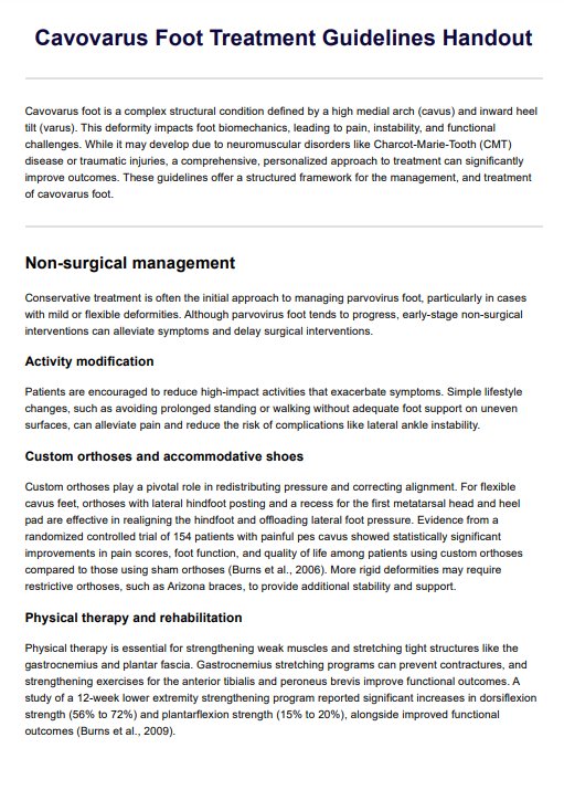
How do healthcare professionals treat cavovarus feet?

The management of cavovarus feet is tailored to relieve symptoms, enhance function, and correct structural abnormalities, with an individualized approach based on the deformity's severity and flexibility. Treatment typically progresses from conservative measures to surgical interventions as needed. This overview highlights five key treatment strategies discussed in clinical guidelines, providing a comprehensive framework for patient care.

Conservative treatments

The first line of management focuses on nonoperative measures designed to alleviate pain, maintain function, and delay the need for surgical intervention.

Orthotic devices, such as custom shoe inserts or arch supports, are critical in redistributing plantar pressure and improving foot alignment. Physical therapy complements orthotic use by targeting specific muscle groups: stretching tight structures like the plantar fascia and Achilles tendon while strengthening weaker muscles, such as the peroneus brevis, to enhance balance and mobility. Anti-inflammatory medications, including non-steroidal anti-inflammatory drugs (NSAIDs), are often prescribed to manage pain and swelling.

While conservative treatments cannot correct the underlying deformity, they provide significant symptom relief, particularly for mild or flexible cases, as supported by clinical studies.

Soft-tissue release

When conservative methods fail to provide adequate relief and the deformity remains flexible, soft-tissue release becomes a key intervention. Procedures such as plantar fasciotomy or Achilles tendon lengthening are employed to alleviate contractures and improve range of motion. These minimally invasive techniques focus on restoring flexibility and preparing the foot for further corrective measures. Soft-tissue release is frequently combined with other interventions, such as tendon transfers or bony reconstruction, to optimize outcomes and prevent recurrence.

Soft tissue balancing techniques

Tendon transfers are particularly effective in addressing muscle imbalances and dynamic deformities associated with cavovarus feet. These procedures involve redirecting tendons to new positions to restore functional balance:

  • Posterior tibial tendon transfer: Relocates the tendon laterally to counteract hindfoot varus and reduce inward heel rolling.
  • Peroneus longus to brevis transfer: Addresses forefoot imbalances by reducing plantarflexion forces on the first ray and enhancing lateral stability.

Tendon transfers are most successful when the deformity is flexible, and the affected muscles retain adequate strength to adapt to their new roles. They are often combined with soft tissue releases or bony reconstruction for comprehensive correction.

Bony reconstruction

In severe or rigid deformities, bony reconstruction is required to correct structural misalignments and restore a plantigrade, functional foot. Techniques include:

  • Osteotomies: Procedures such as dorsiflexion wedge osteotomies for forefoot deformities and calcaneal osteotomies for hindfoot realignment reshape and reposition bones to correct angular deformities.
  • Arthrodesis: Joint fusion procedures, including double or triple arthrodesis, are reserved for cases with significant joint damage or arthritis, stabilizing the foot while eliminating pain.

Bony reconstruction is highly individualized, with the surgical plan guided by imaging studies and the deformity's characteristics. For complex cases, a combination of osteotomies and arthrodesis may be required to achieve optimal outcomes.

Comprehensive, personalized care

Treatment of cavovarus feet is most effective when guided by a personalized approach that considers the patient’s functional needs, deformity severity, and underlying etiology. By combining these strategies—conservative management, soft-tissue release, tendon transfers, and bony reconstruction—healthcare professionals can achieve significant improvements in pain relief, alignment, and mobility, tailoring interventions to the unique challenges posed by each patient’s condition.

How to use our Cavovarus Foot Treatment Guidelines Handout

Our treatment guidelines handout template for cavovarus foot is designed to provide a clear and structured approach to managing this complex foot condition. Follow these steps to make the most of the handout in your practice:

Step 1: Download the template

Access the Treatment Guidelines for Cavovarus Foot Handout by clicking "Use template" for a version you can edit via the Carepatron app. For a PDF copy, choose "Download."

Step 2: Use in clinical practice

Refer to the handout as a comprehensive guide when evaluating and managing patients with cavovarus foot. It includes evidence-based treatments, ranging from conservative options such as orthotics and physical therapy to surgical interventions like tendon transfers and osteotomies.

Step 3: Share with patients and caregivers

Provide the handout to patients and their caregivers to help them understand their condition and the available treatment options. This fosters patient engagement and allows them to make informed decisions about their care plan.

Step 4: Keep for future reference

Store a copy of the handout in the patient’s file. This ensures continuity of care by serving as a quick reference during follow-up visits and facilitating collaboration among healthcare providers involved in the patient’s treatment.

How healthcare professionals will benefit from this handout

Healthcare professionals will benefit from our cavovarus foot treatment handout by gaining a streamlined and comprehensive resource that supports evidence-based care. The handout consolidates key treatment strategies, ranging from conservative approaches to advanced surgical options, providing a clear framework for managing this complex foot deformity. This structure enables clinicians to make informed decisions tailored to the severity and etiology of the patient’s condition, ensuring a personalized approach to treatment.

By using the handout, professionals can save time during consultations, as it serves as a quick reference for management approaches. It also helps standardize care across providers, promoting consistency in treatment planning and reducing the risk of oversight. Furthermore, the handout can be shared with patients and caregivers, improving communication and fostering a collaborative approach to care. Patients who understand their condition and the rationale behind their treatment are more likely to adhere to recommendations, enhancing clinical outcomes.

The handout also facilitates interdisciplinary collaboration by providing a common resource for all team members involved in the patient’s care. By leveraging this tool, healthcare professionals can enhance efficiency, patient engagement, and the overall quality of care for individuals with cavovarus foot.

Reference

Qin, B., Wu, S., & Zhang, H. (2022). Evaluation and Management of Cavus Foot in Adults: A Narrative Review. Journal of clinical medicine, 11(13), 3679. https://doi.org/10.3390/jcm11133679

How is a cavus foot diagnosed?

Commonly asked questions

How is a cavus foot diagnosed?

A cavovarus foot deformity is diagnosed through a combination of clinical evaluations, imaging, and medical history. Healthcare professionals assess the structure of the foot, looking for signs such as a high arch, heel bone misalignment, or foot weakness. Tests like the Coleman block test can determine the flexibility of the deformity, while X-rays, CT scans, or MRIs provide detailed imaging of the ankle joint and foot structure.
What conservative treatments are available for a cavus foot deformity?

Conservative treatments aim to alleviate symptoms such as foot pain and instability. Physical therapy is commonly recommended to strengthen weak muscles and improve balance, while orthotic shoe wear can provide support for high arch feet and redistribute pressure. Stretching the plantar fascia and avoiding activities that exacerbate ankle sprains can also help manage the condition without surgery.

When is surgery necessary for cavus foot, and what are the options?

Cavus foot surgery is typically recommended when conservative treatments fail to relieve symptoms, or the deformity causes significant functional issues. Surgical options include soft tissue procedures, such as plantar fascia release or tendon transfer, to rebalance muscle forces. For more severe cases, osteotomies or joint fusion may be performed to realign the heel bone and correct the high arch.

Join 10,000+ teams using Carepatron to be more productive

One app for all your healthcare work