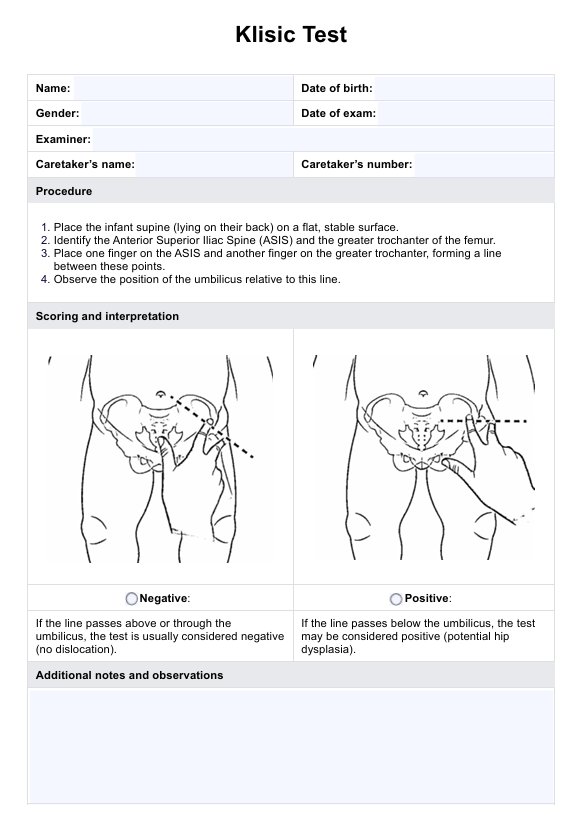
Knee Anatomy Diagram
Access our free Knee Anatomy Diagram to enhance communication, improve patient education, and support accurate diagnosis and treatment planning.

Fact Checked by Karina Jimenea.


What is a Knee Anatomy Diagram?
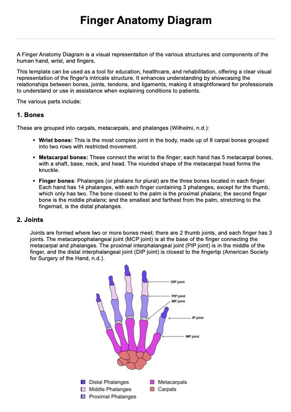
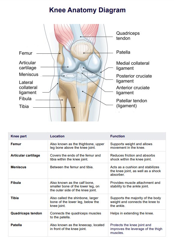
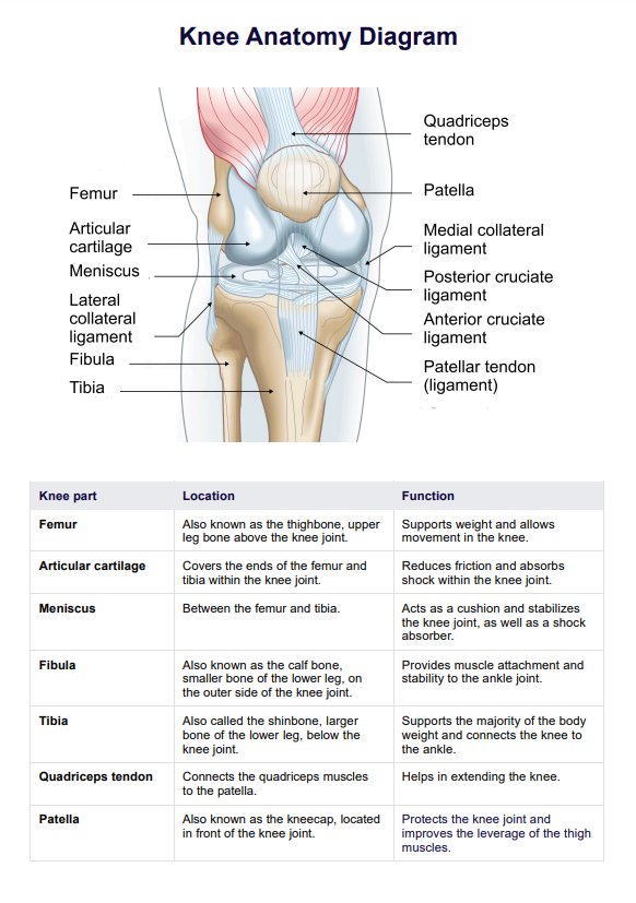
Our Knee Anatomy Diagram is an illustrated representation of the knee joint and its surrounding structures. It highlights the location, structure, and connections of bones, ligaments, tendons, and muscles within the knee.
This diagram is valuable for healthcare professionals, such as physical therapists, orthopedic specialists, and sports medicine practitioners. It allows them to explain the knee's anatomy and function more effectively to patients.
Knee Anatomy Diagrams provide a visual aid that enhances patient comprehension and improves treatment approaches for knee injuries. They are also widely used in educational settings for anatomy, physiotherapy, or sports science students.
Knee Anatomy Diagram Template
Knee Anatomy Diagram Sample
How to use our Knee Anatomy Diagram template
This handout is designed to help you effectively communicate and educate patients about knee anatomy and function. Follow these steps to maximize its utility:
Step 1: Access the handout
Open the hadout via the Carepatron app by clicking the "Use template" button. You can also download a PDF version by choosing "Download."
Step 2: Review and customize
Familiarize yourself with the diagrams and content to ensure clarity and accuracy during consultations. Add annotations or highlights specific to your specialty or a patient’s condition to tailor the handout for more personalized education.
Step 3: Use as a consultation tool
Leverage the handout as a visual reference to simplify complex explanations of knee anatomy, function, and common injuries. Highlight specific ligaments, bones, or muscles related to conditions or symptoms being discussed. Enhance patient understanding and engagement with clear, structured visuals.
Step 4: Provide as an educational resource
When applicable, give the handout to patients post-consultation to reinforce their understanding of their condition, injury, or treatment plan. This ensures they have a reliable, professional resource for future reference.
Benefits of using this diagram
A Knee Anatomy Diagram represents the knee’s complex structure, including bones, ligaments, tendons, muscles, and cartilage. The diagram illustrates how these components interact and helps healthcare professionals and patients better understand knee mechanics, injury locations, and functional movements during consultations. Here are the key advantages of using our template:
Improved understanding
The Knee Anatomy Diagram offers a straightforward way to visualize the knee's structure, helping you and your patients accurately identify key components and their functions. By clearly mapping out how bones, ligaments, and muscles connect, the diagram aids in explaining movement, stability, and areas of concern during patient sessions.
Enhanced patient communication
Effective communication is essential in patient care. A Knee Anatomy Diagram simplifies complex explanations, making discussing conditions such as ligament tears, meniscus injuries, and joint degeneration easier. Patients gain a more precise visual understanding of their diagnosis, reducing confusion and improving adherence to rehabilitation plans.
Better treatment planning
You can better illustrate treatment options, surgical procedures, or rehabilitation strategies using a visual reference. This helps patients make informed decisions about their care, leading to more effective recovery and long-term joint health.
Common knee injuries and their impact on knee function
The knee joint is a hinge joint that supports body weight and allows knee flexion and extension. It consists of three bones—the femur (thigh bone), tibia (shin bone), and patella (kneecap)—along with crucial knee ligaments, tendons, cartilage, and connective tissues that provide knee stability and mobility. Due to its complex structure and weight-bearing role, the knee is vulnerable to various injuries, including ligament tears, tendon strains, and cartilage damage. We've listed some below to help you out:
Ligament injuries
Ligaments are connective tissue structures that stabilize the knee and prevent excessive movement. Damage to these ligaments can significantly affect knee stability and function.
- Anterior cruciate ligament (ACL) injury: Often caused by sudden stops, pivots, or direct impact, ACL tears commonly occur in sports. This injury affects knee stability and can lead to joint effusion (swelling due to synovial fluid accumulation).
- Posterior cruciate ligament (PCL) injury: The PCL, located at the posterior aspect of the knee, is injured due to valgus stress, hyperextension, or direct impact. It primarily affects posterior tibial movement and knee flexion.
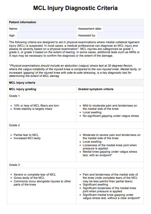
- Medial collateral ligament (MCL) injury:The MCL stabilizes the medial aspect of the knee. Injury often results from valgus stress (force pushing the knee inward), leading to joint capsule and soft tissue damage.
- Lateral collateral ligament (LCL) injury: The LCL stabilizes the lateral aspect of the knee and is injured due to excessive outward force, affecting lateral knee stability.
Cartilage and meniscus injuries
Cartilage and menisci act as shock absorbers between the knee's articulating surfaces. Damage can lead to pain, swelling, and reduced mobility.
- Medial and lateral meniscus tears: The medial meniscus and lateral meniscus are crescent-shaped shock absorbers located on the tibial plateau. They aid in weight distribution and knee stability. Twisting motions, especially during weight-bearing activities, can cause meniscus tears, leading to pain and joint instability.
- Articular cartilage damage: Articular cartilage covers the femoral condyles, medial condyle, lateral condyle, and tibial condyles. Injury to this cartilage can result in joint degeneration, pain, and impaired movement.
Tendon injuries
Tendons connect muscles to bones, enabling movement and absorbing stress. Overuse, trauma, or degeneration can lead to tendon injuries.
- Quadriceps tendon injury: The quadriceps tendon connects the quadriceps muscle to the patella and is essential for knee extension. Strains or tears can make straightening the knee difficult.
- Patellar tendonitis (jumper’s knee): The patellar tendon links the patella to the proximal tibia, playing a crucial role in knee flexors and extension. Repetitive jumping or running can cause inflammation, leading to pain and limited movement.
Other knee conditions
- Joint effusion (water on the knee): Excess synovial fluid accumulation due to injury or inflammation leads to swelling, stiffness, and pain.
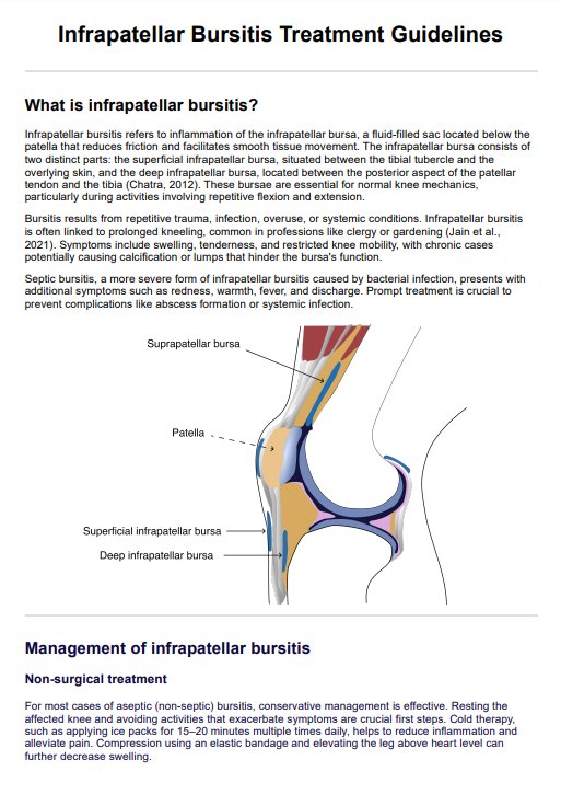
- Bursitis: Inflammation of the fluid-filled sacs around the knee, particularly the joint capsule, causes pain and restricted movement.
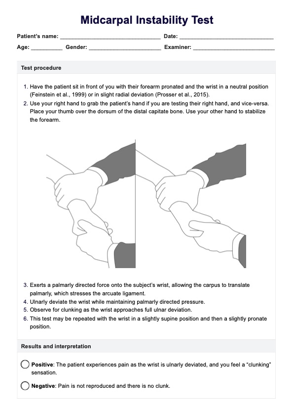
- Knee instability: Damage to knee ligaments, the knee joint capsule, or the tibial nerve can result in instability, making the knee move improperly or feel weak during weight-bearing activities.
Using a Knee Anatomy Diagram alongside your assessment makes spotting and explaining these common injuries much easier. It’s a simple way to improve diagnosis and treatment planning and help patients understand what’s going on with their knee.
Commonly asked questions
The knee has two femoral condyles—one on the inside and one on the outside. The medial femoral condyle is situated on the inner side of the knee, while the lateral femoral condyle, which is larger, is found on the outer side. These condyles play a key role in the knee’s overall movement, supporting the joint during bending and providing a smooth surface for the femur to glide over the tibia.
A Knee Anatomy Diagram provides a clear, structured visual representation of the knee's components, including bones, ligaments, tendons, muscles, and vascular structures. It enhances patient education by simplifying complex anatomical concepts, allowing healthcare providers to explain injuries, conditions, and treatment plans in a way that is easy for patients to understand.
The diagram is a valuable diagnostic tool because it clearly illustrates the locations of key anatomical structures, such as the patellar ligament, medial tibial plateau, and femoral condyles. This helps healthcare professionals identify injury sites, assess stability, and develop targeted rehabilitation or surgical plans, ensuring more effective treatment strategies.
A Knee Anatomy Diagram highlights the knee as a synovial joint, emphasizing the synovial membrane's and fluid's role in reducing friction between the articular surfaces. It also depicts the knee’s function as a weight-bearing joint.
Related Templates

Popular Templates