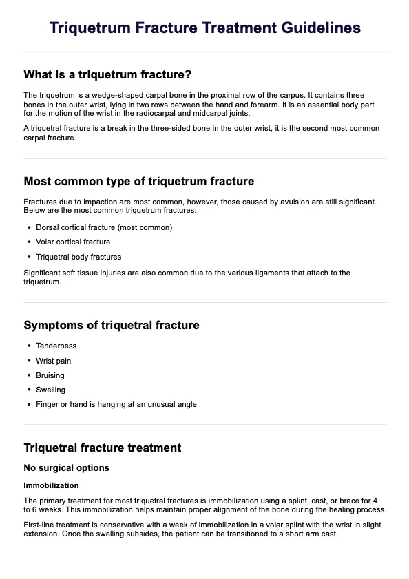
Triquetrum Fracture Treatment Guidelines
Click here to learn more about a triquetrum fracture. Download a free Triquetrum Fracture Treatment Guidelines for better patient education.
What is a triquetrum fracture?
The triquetrum is a wedge-shaped carpal bone in the proximal row of the carpus (Guo et al., 2021). It contains three bones in the outer wrist, lying in two rows between the hand and forearm. It is an essential body part for the motion of the wrist in the radiocarpal and midcarpal joints (Nanduri et al., 2022).
A triquetral fracture is a break in the three-sided bone in the outer wrist; it is the second most common carpal fracture (Guo et al., 2021). The protection of the triquetrum from surrounding carpal bones means that carpal bone fractures are typically caused by the impact of adjacent bony structures or the avulsion of attached ligaments rather than direct force. Fractures due to impaction are the most common. However, those caused by avulsion are still significant (Guo et al., 2021).
The most common cause of a triquetral fracture is a fall onto an outstretched hand with the wrist in ulnar deviation or extension. The typical mechanisms are impaction by the hamate or ulnar styloid, and avulsion from the dorsal radiotriquetral or dorsal scaphotriquetral ligaments.
The dorsal cortical fracture is the most common within triquetral fractures, accounting for 93-95% of triquetral fractures (Nanduri et al., 2022). The second and third most common types are volar cortical and triquetral body fractures (Guo et al., 2021). It is also common for significant soft tissue injuries to occur due to the various ligaments that attach to the triquetrum, commonly the dorsal scaphotriquetral ligament (Becce et al., 2013).
The main symptoms of a triquetral fracture are tenderness and wrist pain. There also may be bruising and swelling, or you may notice your finger or hand hanging at an unusual angle. In addition, there may be pain when you attempt to grip something, bend your wrist, or make a fist.
Triquetrum Fracture Treatment Guidelines Template
Triquetrum Fracture Treatment Guidelines Example
Triquetral fracture treatment
Treatment aims to restore wrist function and prevent complications like chronic pain or instability. The management approach depends on the severity and displacement of the fracture.
Non-surgical treatment
Typically, triquetral fractures can be fixed through non-surgical treatments, particularly nondisplaced and dorsal cortical fracture patterns (Guo et al., 2021). Immobilization in a volar splint with the wrist in slight extension for a week is the first-choice treatment (Guo et al., 2021).
After swelling decreases, it is common to move the patient to a short arm cast and, at four weeks, transition to a removable wrist splint (Guo et al., 2021). After eight weeks out of the brace, patients should be able to resume normal activities (Guo et al., 2021). The majority of triquetral fractures respond favorably to immobilization for 4-6 weeks (Nanduri et al., 2022).
Surgical treatments
Surgical treatments should only be considered in fractures with significant displacement and clinical concern for fracture dislocation or carpal instability (Nanduri et al., 2022). In this case, the fracture fragment can be surgically removed.
For more detailed treatment options, access our treatment guidelines handout template.
How to use our treatment guidelines handout template?
Our Triquetrum Fracture Treatment Guidelines handout is free and easy to use. Follow these steps to get started:
Step 1: Download the template
Access the Triquetrum Fracture Treatment Guidelines template by clicking "Use template," allowing you to edit the resource via the Carepatron app. For a PDF copy, choose "Download."
Step 2: Familiarize yourself with the template
Review the Triquetrum Fracture Treatment Guidelines template to understand its structure and content. The template is structured to guide you through what a triquetral fracture is, the most common types of fracture, and its associated injuries. It further describes the common causes and symptoms, along with treatment guidelines.
Step 3: Use the template during consultations
Follow the template when discussing the patient's triquetrum fracture. It will help ensure you provide clear and comprehensive details so the patient has the correct and necessary information to understand their treatment plan following their injury.
Step 4: Give a copy to patients
Share the Triquetrum Fracture Treatment Guidelines template with the patient to reinforce the information discussed. This allows them to review the details at their own pace.
How can healthcare professionals benefit from our template?
The Triquetrum Fracture Treatment Guidelines template offers healthcare professionals a clear, evidence-based framework for managing wrist injuries. It streamlines common causes, symptoms, and treatment options based on fracture severity and ensures consistent, high-quality care.
The template saves time during consultations by providing quick, structured guidance and fosters better collaboration between clinicians, radiologists, and patients through standardized protocols. It also serves as a valuable educational tool for less experienced practitioners while offering a reliable checklist for seasoned professionals.
References
Becce, F., Theumann, N., Bollmann, C., Omoumi, P., Richarme, D., Guerini, H., ... & Drapé, J. L. (2013). Dorsal fractures of the triquetrum: MRI findings with an emphasis on dorsal carpal ligament injuries. American Journal of Roentgenology, 200(3), 608-617.
Guo, R. C., Cardenas, J. M., & Wu, C. H. (2021). Triquetral fractures overview. Current Reviews in Musculoskeletal Medicine, 14(2), 101–106. https://doi.org/10.1007/s12178-021-09692-w
Nanduri, A., Kim, A., Nolan, C., Dubey, J., & Barbera, A. (2022). Triquetrum fracture with Pisiform dislocation. Orthopedic Reviews. Retrieved December 09, 2024, from https://orthopedicreviews.openmedicalpublishing.org/article/32339-triquetrum-fracture-with-pisiform-dislocation
Commonly asked questions
A triquetrum fracture is a break in the triquetrum, one of the eight small carpal bones on the ulnar side of the wrist near the pinky finger. As one of the most common carpal fractures, it typically occurs due to a fall on an outstretched hand, direct trauma, or repetitive stress. There are three main types of triquetrum fractures: dorsal cortical fractures, which are the most common and involve a break on the dorsal surface of the bone; body fractures, which affect the main portion of the triquetrum and are often associated with high-impact trauma; and volar cortical fractures, which are less common and occur on the volar (palmar) side of the bone, often linked to ligamentous injury or extreme wrist extension.
A diagnosis is typically made through a physical examination to assess pain and range of motion. Imaging studies, such as X-rays, CT scans, or MRIs, are also done to confirm the fracture and check for associated injuries.
Triquetrum fractures are typically treated with immobilization using a splint or cast to allow the bone to heal. Most cases resolve without complications, but severe or displaced fractures may require surgical intervention. Surgery may involve fixation techniques such as pins or screws to restore stability and alignment.