Suele notarse en la cara interna de la rodilla o en la parte anterior de la articulación de la cadera.
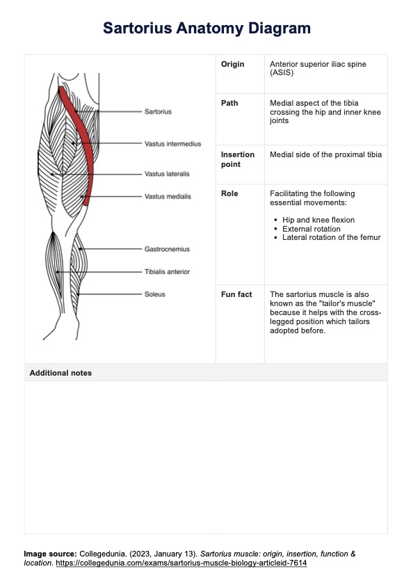
Obtenga una copia de nuestro Diagrama de anatomía del sartorio para beneficiarse de sus usos educativos y clínicos, incluido el dominio de la anatomía del músculo sartorio.

Suele notarse en la cara interna de la rodilla o en la parte anterior de la articulación de la cadera.
La lesión más común es la distensión.
El tratamiento puede incluir reposo, hielo y el uso de un vendaje compresivo.
EHR and practice management software
*No credit card required
Free
$0/usd
Unlimited clients
Telehealth
1GB of storage
Client portal text
Automated billing and online payments