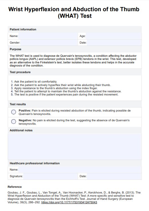
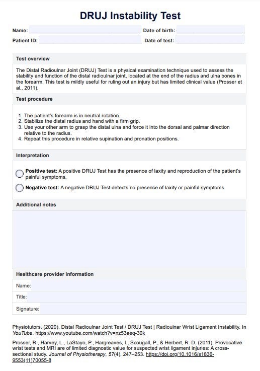
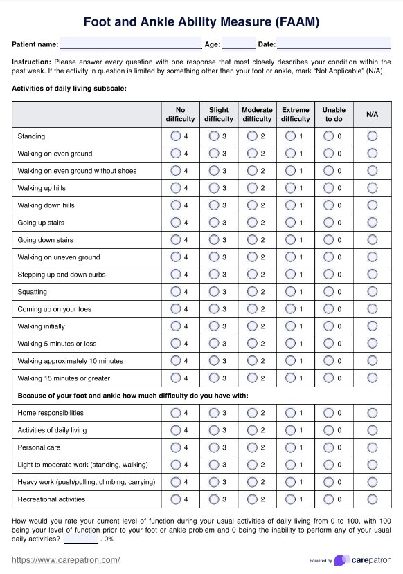
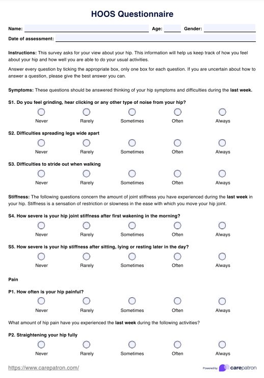
Myotomes Chart
Use our Myotomes chart as a reference for myotomes and muscle groups—for better diagnosis of neuromuscular disorders and spinal nerve injuries.


What is a Myotomes Chart?
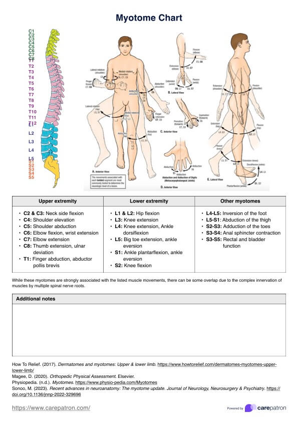
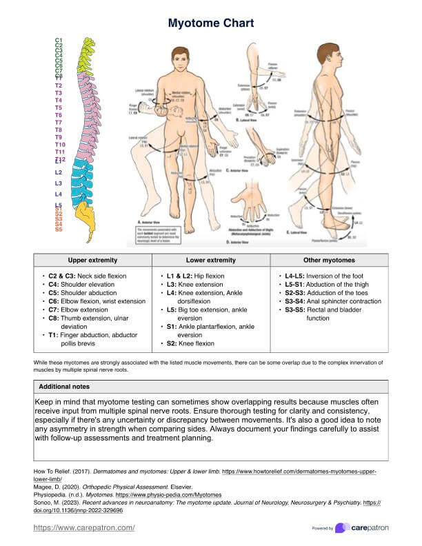
Myotomes refer to the group of muscles innervated by a single spinal nerve root in the peripheral nervous system. Each myotome corresponds to a specific nerve root, which controls muscle movement and function in different parts of the body. A Myotomes Chart visually illustrates this relationship—between specific spinal nerve roots and the muscles they innervate. It is a valuable tool for healthcare professionals, such as doctors, physical therapists, chiropractors, and students, to better understand and diagnose neurological conditions, spinal nerve injuries, and musculoskeletal disorders.
Myotomes Charts are often used in clinical settings to assess the integrity of spinal nerves during physical examinations. By testing muscle strength and range of motion in specific body areas, healthcare providers can determine if there are spinal cord lesions, nerve root compression, or damage. This information can then be used to guide appropriate treatment strategies and monitor progress during rehabilitation.
A Myotomes Chart can also enhance the understanding of neuromuscular anatomy and physiology among healthcare practitioners. This knowledge can be particularly beneficial when working with patients who have experienced spinal cord injuries, strokes, or other neurological conditions that affect muscle function.
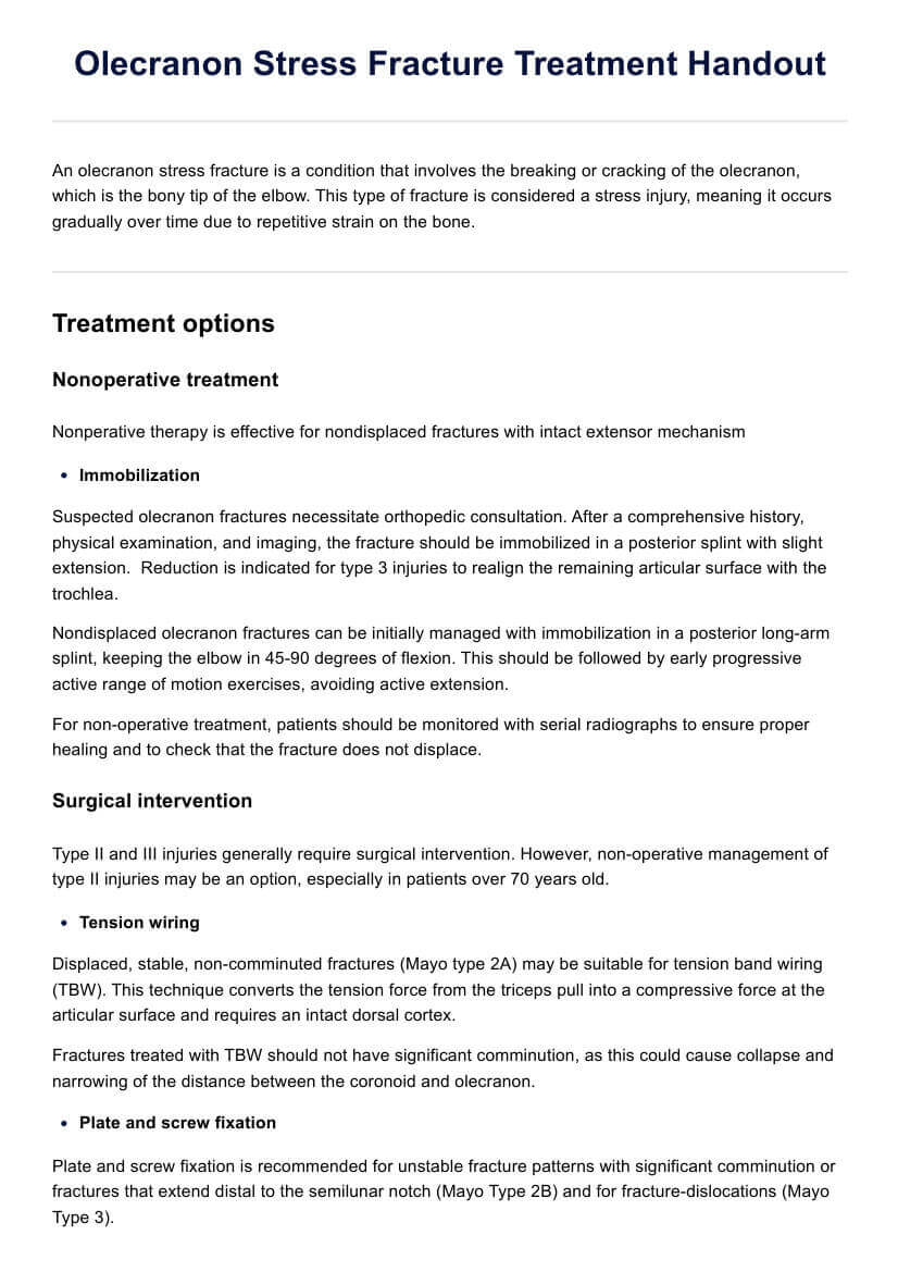
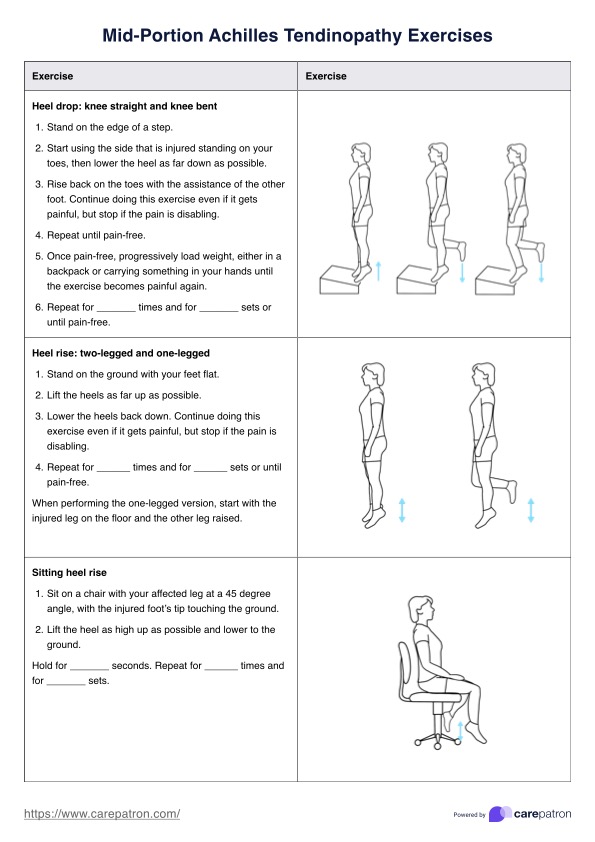
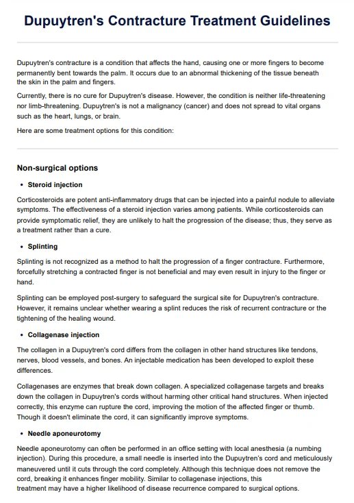
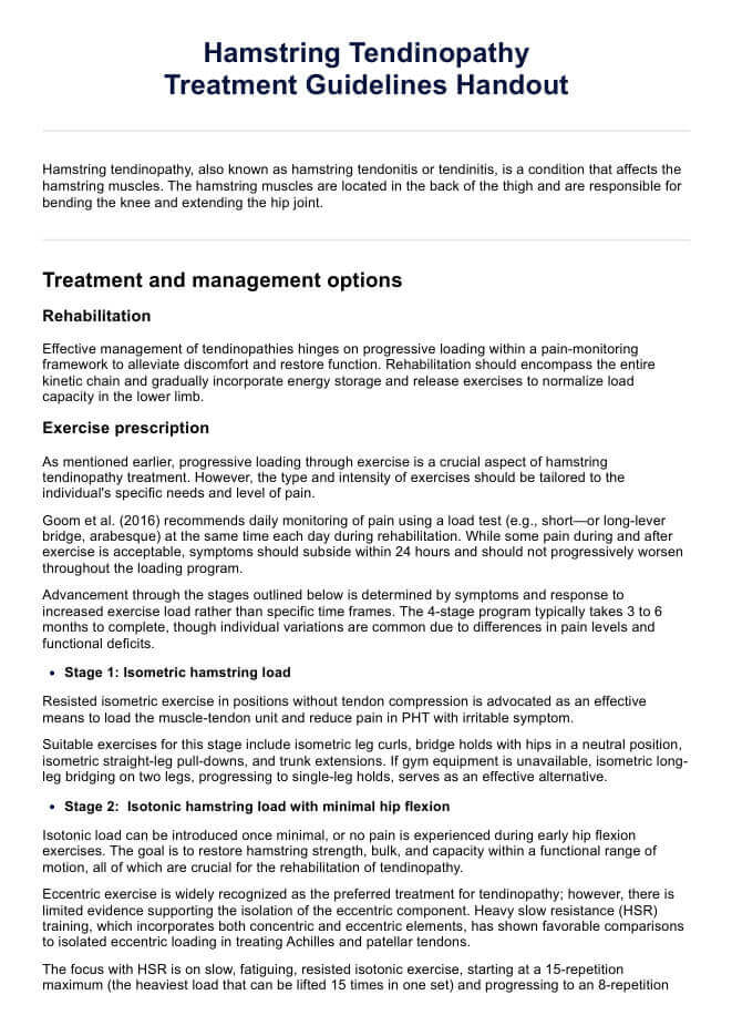
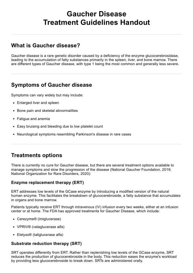
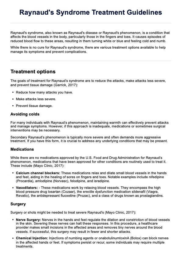
Myotomes Chart Template
Myotomes Chart Example
How to use our Myotomes Chart template
Our printable Myotomes Chart template is valuable for healthcare professionals to assess and diagnose neurological conditions and spinal nerve injuries. Here's how to use it:
Step 1: Access the template
Click "Use Template" to open it in the Carepatron app, where you can customize the template before printing or sharing it. You can also click "Download" for a non-customizable PDF version that can be filled out digitally or printed.
Step 2: Use the template as a reference
Use the Myotomes Chart as a reference during clinical assessments. Whether testing a patient's muscle strength or performing neurological exams, you can quickly identify which spinal nerve is associated with specific muscle movements.
Step 3: Make copies for clinical use
Print or post copies of the Myotomes Chart in your practice for quick reference during patient evaluations. You can also distribute copies to patients or students for educational purposes.
Step 4: Educate patients and students
If you're working with medical students or explaining neuromuscular conditions to patients, the chart can serve as a visual aid. It helps clarify how spinal nerves control muscle function and aids in explaining the effects of spinal injuries or neurological conditions.
Step 5: Monitor and update the care plan
Use the Myotomes Chart as part of a larger care plan to monitor patient progress, evaluate treatment efficacy, and update interventions as necessary.
What is in the template?
The Myotomes Chart template contains a detailed list of myotomes and muscle groups or movements associated with each spinal nerve root. It highlights critical movements like elbow flexion, hip flexion, knee extension, and thumb extension, alongside their corresponding myotomes.
Here's an overview of the movements and the related body parts:
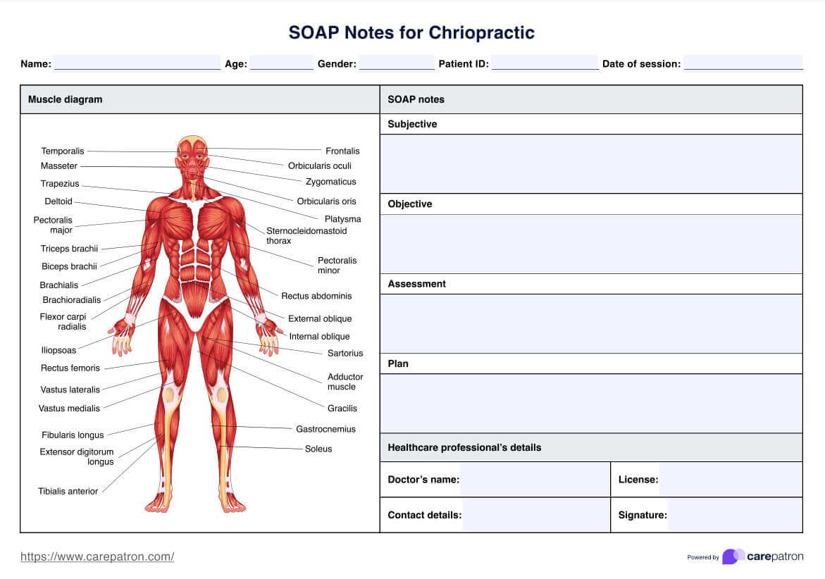
- Elbow flexion: Controlled by the C5 and C6 spinal nerve roots, this movement primarily involves the biceps brachii and brachialis muscles, which allow the bending of the elbow. Additionally, the extensor carpi radialis longus and extensor carpi radialis brevis assist in wrist extension, while flexor carpi radialis helps in wrist flexion.
- Shoulder abduction: The C5 nerve root is responsible for shoulder abduction, involving the deltoid muscle, particularly its middle fibers. This movement is essential for raising the arm laterally away from the body.
- Thumb extension and ulnar deviation: The C8 nerve root controls thumb extension, facilitated by muscles like the extensor pollicis longus. The C8 also influences the flexor carpi ulnaris for ulnar deviation, moving the hand towards the pinky side of the wrist.
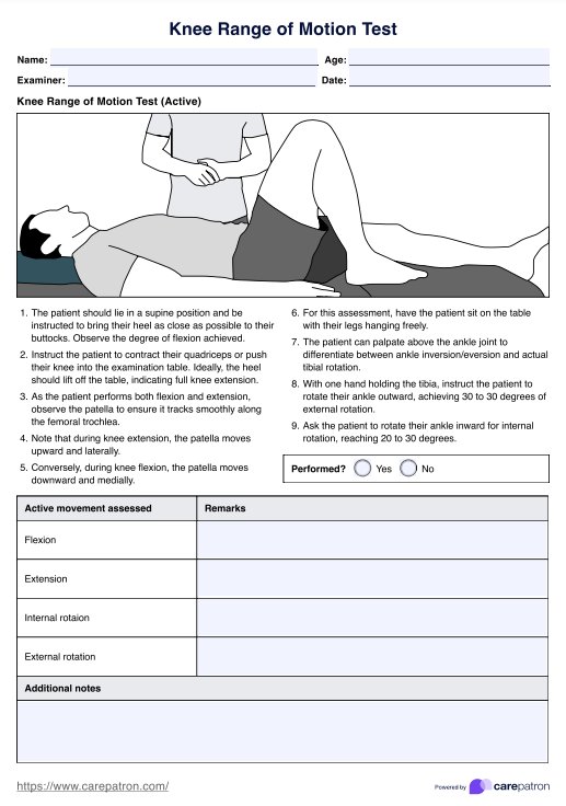
- Knee extension: The L3 and L4 spinal nerve roots control this movement, primarily affecting the quadriceps femoris group, which extends the knee joint. These muscles, crucial for standing and walking, receive innervation from the lumbar myotomes.
- Ankle dorsiflexion: This movement is managed by the L4 nerve root and involves the tibialis anterior muscle, which pulls the foot upward. It's vital for walking, particularly during the swing phase of gait.
- Hip flexion: The L1 and L2 nerve roots innervate muscles like the iliopsoas to allow flexion at the hip joint, a key movement for activities like walking and climbing stairs. The lower limb muscles involved in hip flexion are essential for leg mobility.
- Big toe extension and ankle eversion: The L5 nerve root controls big toe extension through the extensor hallucis longus. Ankle eversion, where the foot moves outward, is managed by the same nerve root, impacting muscles such as the fibularis longus and fibularis brevis.
- Ankle plantarflexion: The S1 nerve root handles this movement, involving the gastrocnemius and soleus muscles, which point the foot downward. This movement is essential for pushing off during walking or running.
- Finger abduction: Controlled by the T1 spinal nerve root, this movement affects the abductor pollicis brevis muscle, which is responsible for spreading the fingers apart. It's a critical movement for hand dexterity and grip strength.
- Knee flexion: The S2 nerve root facilitates knee flexion through the hamstring muscles, crucial for bending the knee and enabling movements such as running or crouching.
When would you use this template?
A versatile resource for healthcare specialists, the Myotomes Chart template is helpful in areas like neurology, physiotherapy, orthopedics, chiropractic interventions, sports medicine, and education.
The template assists in a range of scenarios:
- Neurological assessments: Identifying muscle weaknesses or sensory changes related to spinal nerves, enabling accurate diagnoses and treatments.
- Physiotherapy evaluations: Developing individualized treatment plans targeting affected myotomes and spinal nerves.
- Orthopedic evaluations: Assessing spine-related issues and identifying potential nerve root compression or damage for appropriate treatment plans.
- Chiropractic care: Addressing musculoskeletal pain or dysfunction by understanding spinal nerves and myotomes relationships.
- Sports medicine: Assessing athletes' neuromuscular function and creating targeted rehabilitation programs after injuries or surgeries involving the spine or peripheral nerves.
- Education and training: Simplifying the learning process and improving neuromuscular anatomy and physiology retention for medical students and healthcare professionals.
Commonly asked questions
We test myotomes to identify muscle weakness and assess the function of specific spinal nerve roots. This helps detect neurological disorders or spinal nerve injuries.
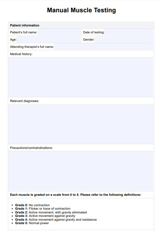
The grading of myotomes typically follows a scale from 0 to 5, where 0 indicates no muscle movement and 5 represents normal muscle strength. This grading helps determine the extent of muscle function loss.
No, dermatome and myotome maps are not the same. Dermatomes refer to skin areas innervated by sensory nerves from a single spinal nerve, while myotomes refer to muscle groups controlled by motor nerves from a single spinal nerve root.


