MCL Anatomy Diagram
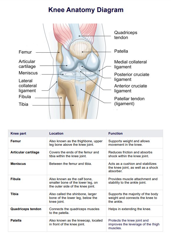
Have a go-to reference of the medial collateral ligament (MCL) for diagnosing or studying by downloading our MCL Anatomy Diagram.


What is the function of the medial collateral ligament (MCL)?
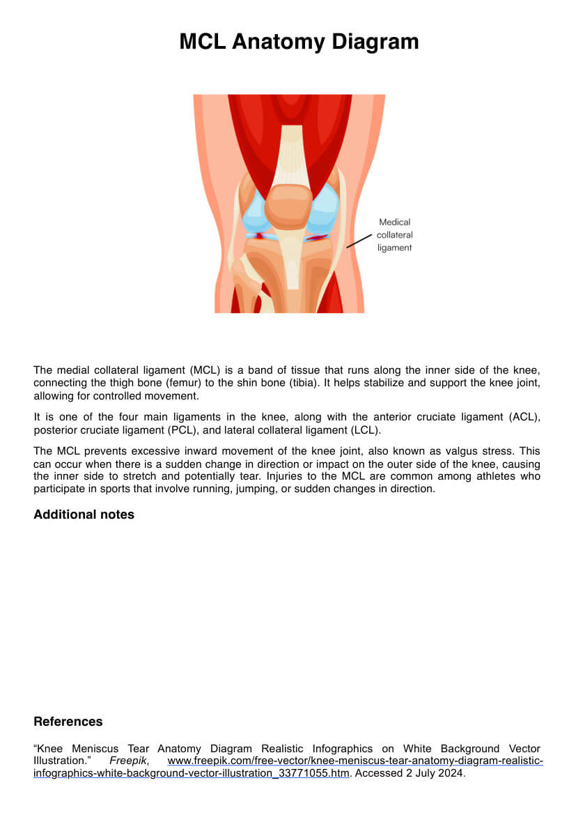
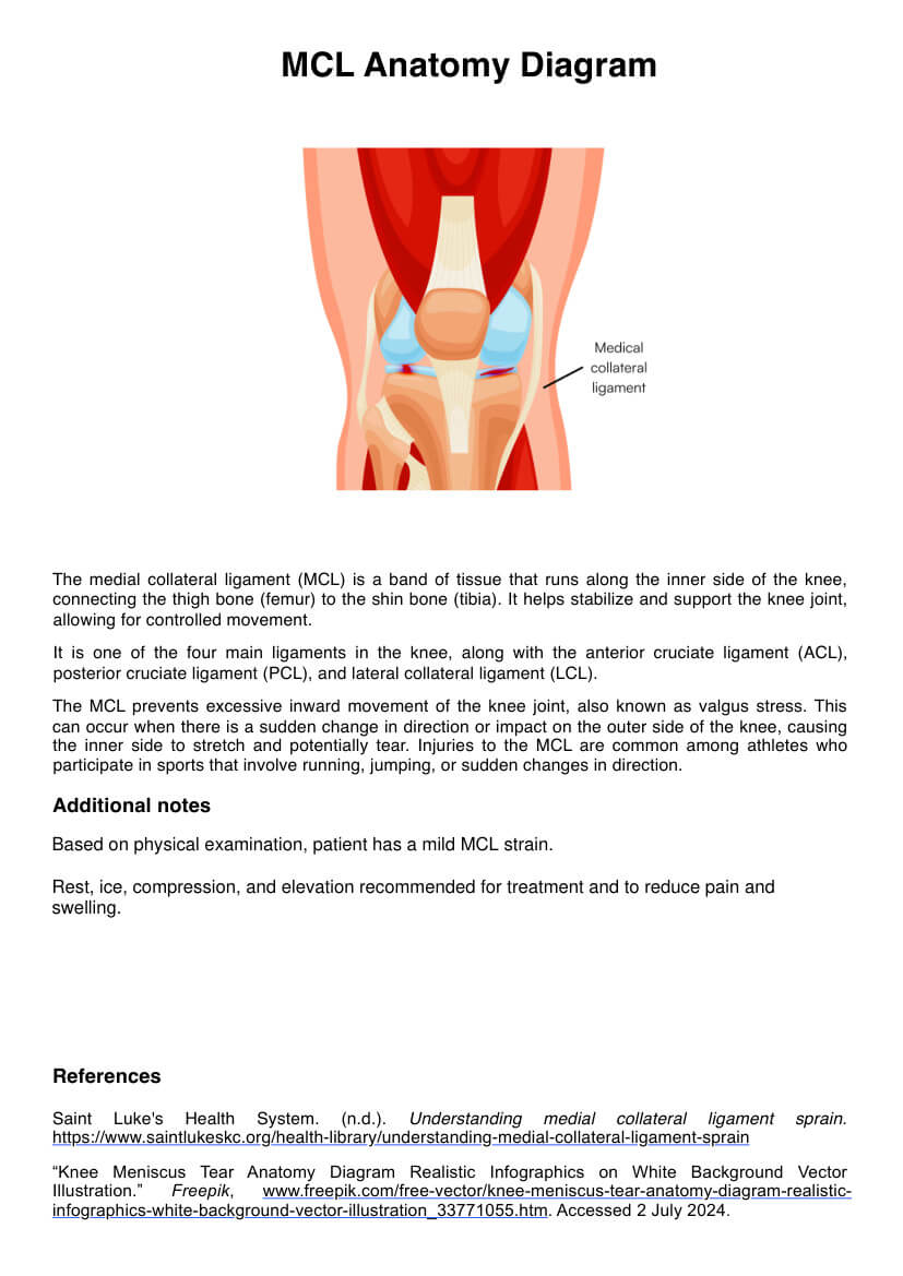
The medial collateral ligament (MCL), also known as the tibial collateral ligament TCL, is a flat connective tissue that runs from the medial femoral epicondyle to the medial condyle of the tibia situated on the medial aspect of the knee joint. It is the largest structure on the medial side, given its length of 8 to 10 cm.
Its primary role is to maintain knee stability by resisting valgus forces, thus preventing the knee from excessively bending inward. In addition, the MCL acts as a primary static stabilizer that assists in the passive stabilization of the knee joint by serving as a secondary restraint to rotational forces and responding to valgus stress.
How is it different from TCLs, ACLs, etc.?
The medial collateral ligament is distinct from other knee ligaments like the anterior cruciate ligament (ACL) and the lateral collateral ligament (LCL), not only in location but also in purpose.
While the ACL is situated deep within the center of the knee, and the LCL is found on the outer side of the knee, the MCL is located on the knee joint’s inner side. Concerning purpose, the MCL’s primary role is to prevent excessive inward bending and to provide rotational stability. This is strikingly different from the ACL, whose primary role is to prevent excessive front-to-back movement, and the LCL’s role is to prevent excessive side-to-side movement, ensuring lateral stability.
Despite these differences, however, these ligaments, along with the posterior cruciate ligament (PCL), posterior oblique ligament (POL), and other ligaments in the knee, contribute to the knee’s overall stability.
What can negatively impact an MCL's functioning?
Given the MCL’s crucial role, one must be aware of the several factors that can impair its function. Primary factors or causes of damage to the MCL are trauma and injuries, often resulting from direct impacts on the outer knee. Examples are cutting maneuvers in sports, such as during a football tackle, awkward landings, or hyperextension in activities like asking. Non-sports-related incidents like car accidents, however, can also damage the MCL.
Aside from these, wear and tear that causes the MCL to lose elasticity, like repetitive stress and activities involving squatting, heavy lifting, and high-impact sports, can also affect its function. Finally, those with previous MCL injuries are also at risk of damage because of an increased likelihood of re-injury.
MCL Anatomy Diagram Template
MCL Anatomy Diagram Sample
What is an MCL Anatomy Diagram?
A MCL Anatomy Diagram is a visual representation of the MCL with its appropriate label plus its corresponding location, which is from the medial epicondyle of the femur (thigh bone) to the medial condyle of the tibia (shin bone), both of which are also located and labeled on the said anatomy diagram.
Like the MCL, the anatomy of the MCL diagram also serves multiple purposes. Healthcare professionals and medical students can incorporate it in their studies as flashcards, educational references for anatomy exams, or even documents where one can note observations and notes during clinical decision-making.
For a copy of an MCL diagram on the knee, access our MCL Anatomy Diagram template by clicking the “Use Template” buttons in this guide or downloading it from our template library. Feel free to save and use it digitally, print it as a hard copy for annotation, or distribute it to patients, students, or colleagues.
Benefits of using this diagram
There are numerous benefits to using a medial collateral ligament (MCL) anatomy diagram. Here’s a list of some of them:
Visual clarity
With the diagram, healthcare professionals and medical students have a clear and precise visual representation of the MCL’s structure, location, and attachments.
Educational aid
Our MCL anatomy diagram simplifies complex anatomical details, making them more accessible and easily understood by medical students, medical professionals, and patients.
Clinical context
Medical professionals can use the diagram during diagnosis to help identify the affected areas and understand the nature of the medial knee injury.
Patient communication
The knee anatomy diagram with the MCL label can facilitate better communication between healthcare providers and patients. With it, patients can understand their condition, including the anatomical basis of their symptoms, and better comprehend treatment options and rehabilitation plans.
Common treatments for Medial Collateral Ligament Injuries
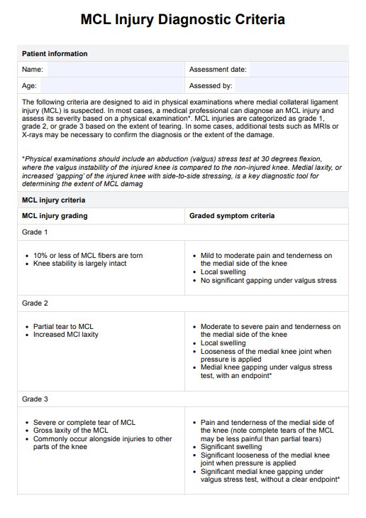
Medial collateral ligament injuries can vary in severity, and treatment is often tailored to the extent of the tear since each grade—Grade 1, Grade 2, and Grade 3—requires different approaches to management and recovery.
Grade 1 MCL tear treatment
For a Grade 1 MCL tear, which is a mild injury, treatment typically involves rest, avoidance of activities that put stress on it, ice to reduce swelling and alleviate pain, and the use of non-steroidal anti-inflammatory drugs (NSAIDs) like ibuprofen for pain management.
As the patient heals, they are encouraged to only gradually return to normal activities when their pain decreases, and mobility improves to promote healing without needing more intensive interventions.
Grade 2 MCL tear treatment
For a moderate tear involving the superficial part of the MCL, a Grade 2 tear, treatment includes crutches and a knee brace to minimize weight-bearing and allow movement while protecting the ligament. NSAIDs or prescribed medications are continued alongside a rehabilitation program to restore function and strength.
Grade 3 MCL tear treatment
As for a Grade 3 MCL tear, which involves the superficial medial collateral ligament and deep medial collateral ligament, the treatment includes rest, immobilization, and, in certain cases, surgery.
Commonly asked questions
The main symptom is pain in the knee right over the ligament. Other symptoms include knee instability, swelling, and bruising.
It depends on the grade. The shortest time is usually a week, and the longest is more than six weeks.
The most common injury is an MCL tear.


