Neuro Eye Exam
A neuro eye exam is a comprehensive assessment of the visual system to diagnose and manage neurological conditions. Download Carepatron's neuro eye exam PDF to learn more about it.


What is a Neuro Eye Exam?
A neuro eye exam is a specialized assessment to evaluate the health and function of the visual system, with a particular focus on the interaction between the eyes and the neurological pathways. Unlike a routine eye exam, which primarily checks for refractive errors and general eye health, a neuro eye exam delves deeper into the intricate connection between the eyes and the brain.
At its core, this examination centers around assessing cranial nerves, which are pivotal in controlling eye movements, pupil responses, and overall visual performance. The cranial nerves, especially the optic nerve, are integral components of the neurological network responsible for transmitting visual information from the eyes to the brain.
Visual acuity, a common term assessed in routine eye exams, is also a vital aspect of the neuro eye exam. However, this examination goes beyond the standard chart reading, exploring nuances of visual function such as depth perception and the ability to perceive spatial relationships accurately. These factors are crucial in understanding how the brain processes and interprets visual information for daily activities.
A neuro eye exam holds particular significance in detecting conditions like cranial nerve palsy, where the normal function of these nerves is compromised. Identifying such issues early on is essential for preventing potential complications and ensuring timely intervention.
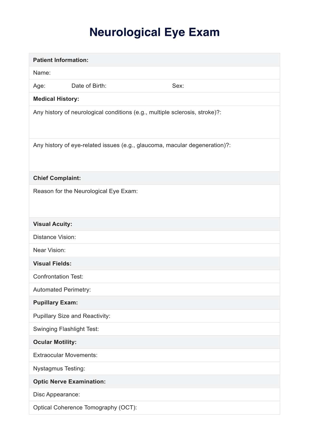
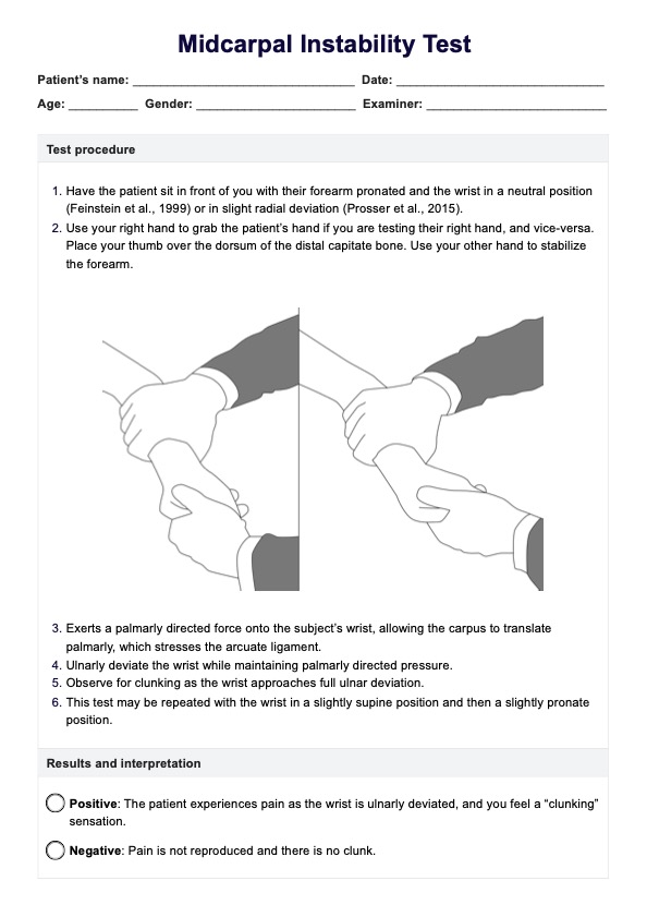
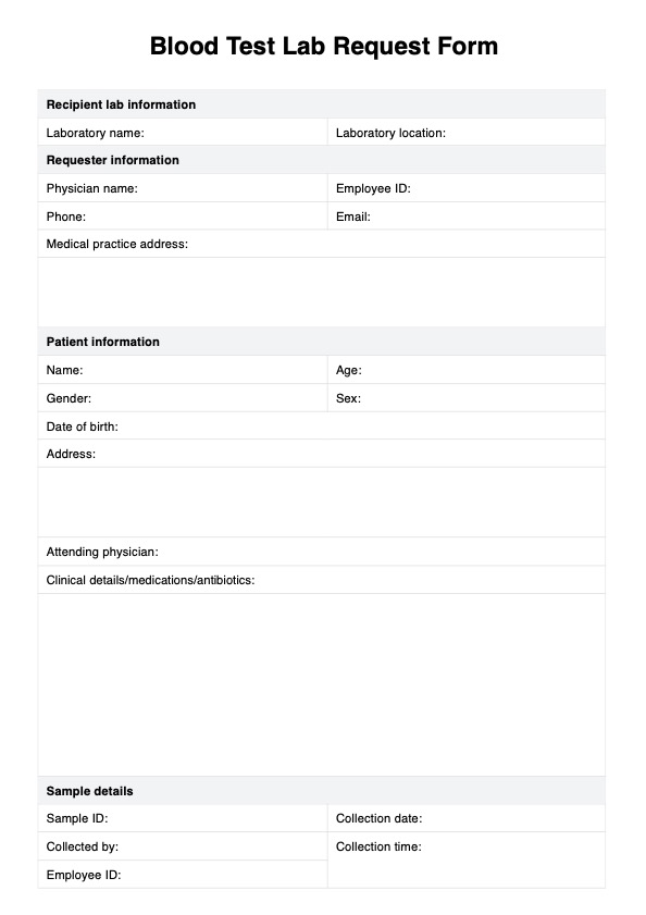
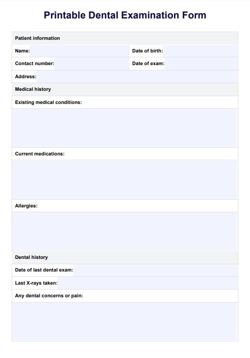
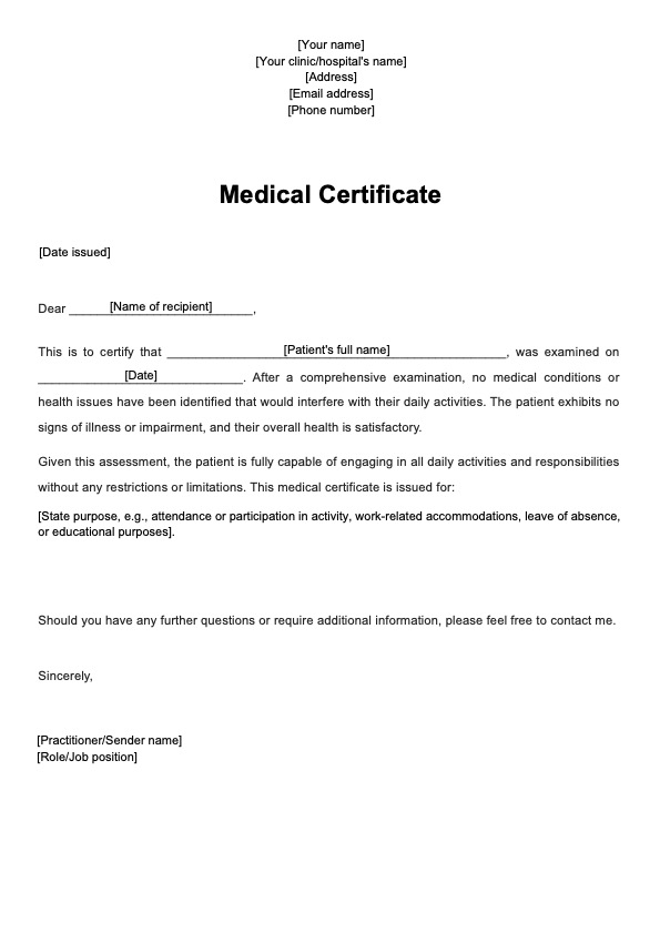
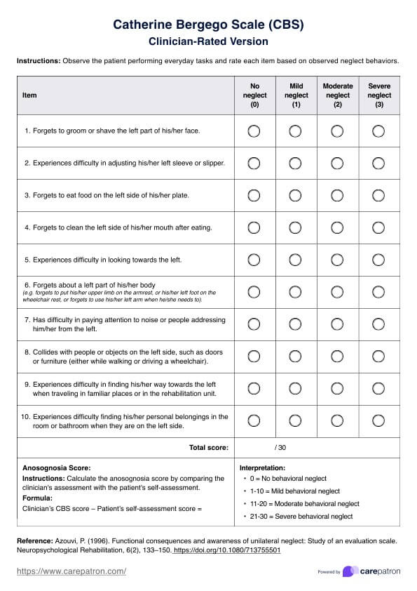
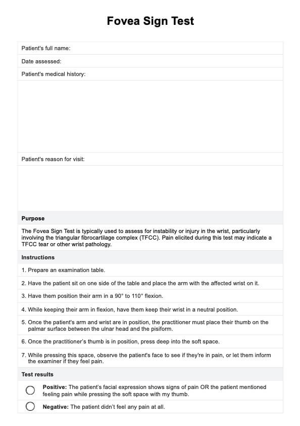
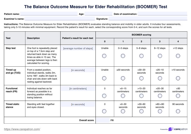
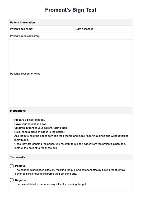
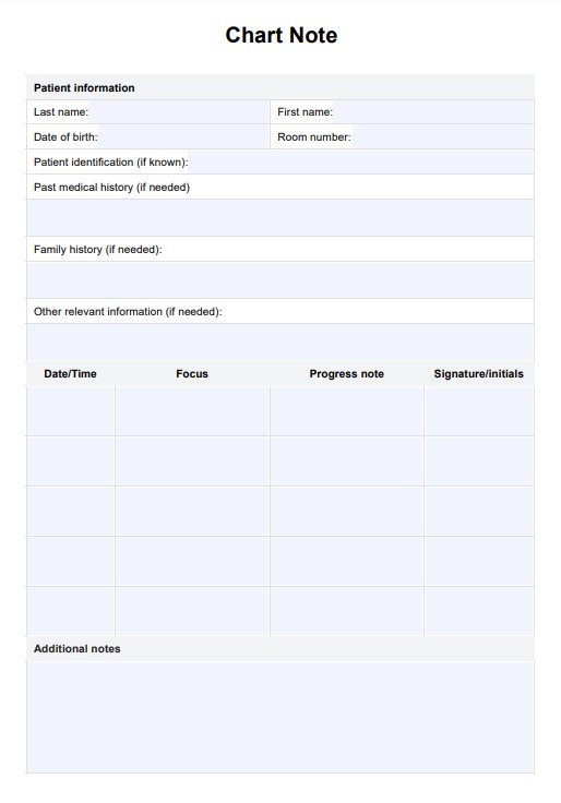
Neuro Eye Exam Template
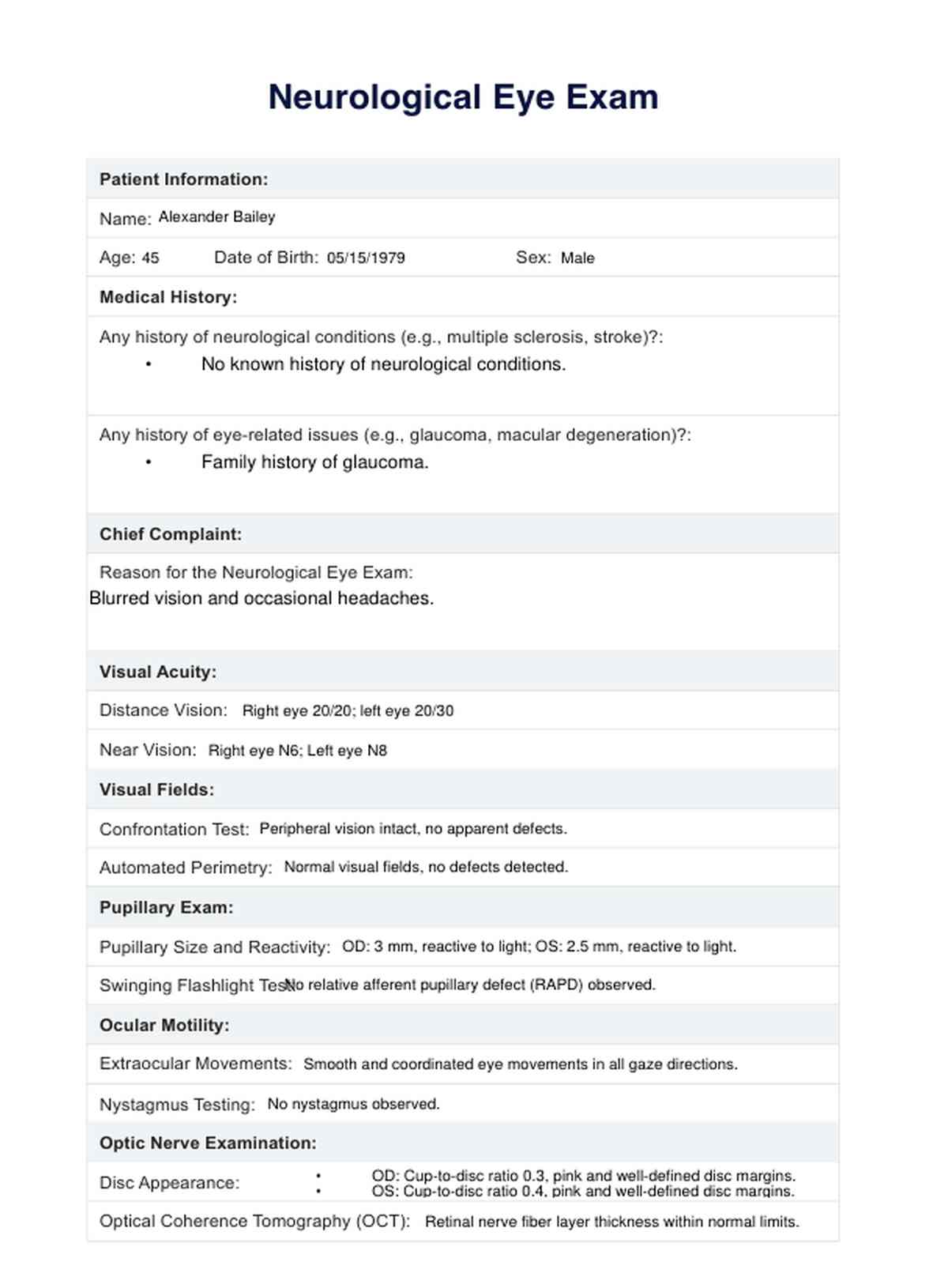
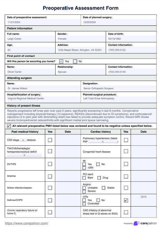
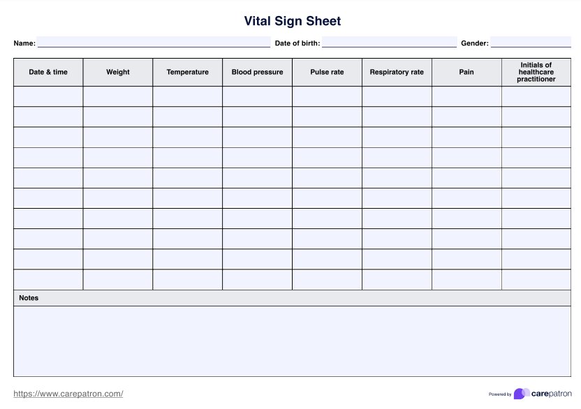
Neuro Eye Exam Example
What conditions does neuro-ophthalmology treat?
Neuro-ophthalmology addresses a diverse range of conditions that involve the intricate interplay between the eyes and the neurological system. This specialized field is equipped to manage various disorders affecting vision and ocular function. Below are some of the conditions that neuro-ophthalmology commonly addresses:
- Optic nerve disorders
- Visual pathway lesions
- Cranial nerve palsy or disorders
- Inflammatory and autoimmune conditions
- Papilledema and increased intracranial pressure
- Double vision or diplopia
- Neurological aspects of ocular mobility disorders
- Pupillary abnormalities
- Neurological implications of ocular conditions
Equipment required for a neuro-ophthalmological assessment
A neuro-ophthalmological assessment requires specialized equipment to thoroughly examine the visual system and its connection to the neurological pathways. Each piece of equipment is crucial in evaluating various eye health and function aspects.
Slit lamp biomicroscope
The slit lamp biomicroscope is an essential tool for examining the anterior and posterior segments of the eye. It aids in assessing the health of the optic nerve and the retina, providing valuable insights into the overall eye structure. This instrument is handy in detecting conditions that may contribute to double vision or impact peripheral vision.
Visual field perimeter
Assessing peripheral vision is integral to a neuro-ophthalmological evaluation. A visual field perimeter helps map the patient's field of vision, detecting any abnormalities that may indicate issues with the eye nerve or other neurological pathways. It is crucial for the differential diagnosis of conditions affecting the visual field.
Pupillometer
A pupillometer is employed to measure the size and reactivity of the pupils. This is essential for identifying a relative afferent pupillary defect (RAPD) associated with asymmetrical pupillary responses and indicative of eye nerve dysfunction. Monitoring pupil responses aids in the comprehensive evaluation of the complete neurological exam.
Diplopia testing tools
Instruments for diplopia testing are crucial for assessing double vision. These tools help determine the nature and extent of diplopia, contributing to a more accurate differential diagnosis. Examining eye movements and coordination with these tools is essential in understanding the neurological basis of double vision.
What does a neurological eye exam include?
A neurological eye exam is a comprehensive assessment designed to evaluate the intricate relationship between the eyes and the neurological system. Here's an overview of what patients can expect from this specialized examination:
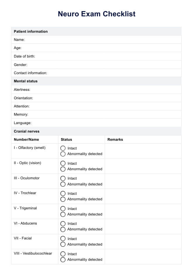
Evaluation of cranial nerves
The neuro-ophthalmologist will thoroughly examine the cranial nerve, particularly the optic nerve. Assessing the functionality of these nerves is crucial, as they play a central role in transmitting visual information from the eyes to the brain. The examination may also involve the trigeminal nerve, contributing to facial sensation and specific eye movements.
Depth perception assessment
Unlike a routine eye exam that primarily checks visual acuity, a neurological eye exam delves into depth perception. This aspect is vital for understanding how the brain interprets spatial relationships and processes three-dimensional visual information.
Visual field testing
Patients can expect to undergo visual field testing to identify any defects or abnormalities in their vision. Detecting visual field defects is essential for diagnosing conditions affecting the optic tract or other neurological pathways.
Optical Coherence Tomography (OCT)
OCT is a non-invasive imaging technique used to capture high-resolution cross-sectional images of the retina. It provides detailed information about the optic nerve and can reveal structural changes that might not be apparent in a routine eye exam. This technology aids in detecting an optic tract lesion and other abnormalities early.
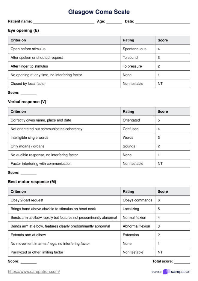
Examination for mental status
Beyond the visual aspects, a neurological eye exam may include an assessment of mental status. This broader evaluation helps understand how neurological conditions may impact vision-related cognitive functions.
Consideration of spinal cord involvement
In some instances, the examination may extend to evaluating the potential involvement of the spinal cord in neurological conditions affecting the eyes. This holistic approach ensures a comprehensive understanding of the patient's neurological health.
How to use the template?
Utilizing the neuro eye exam template effectively involves a systematic approach to ensure a thorough evaluation of the visual system and neurological pathways. Follow these step-by-step guidelines to make the most of the template:
1. Patient information
Begin by introducing the patient and providing relevant demographic information. Include age, gender, and any pertinent medical history that may impact the neurological eye exam.
2. Cranial nerve assessment
This section details the examination of cranial nerves, focusing on the optic and trigeminal nerve. Note any abnormalities or issues observed during the assessment.
3. Depth perception analysis
Explore the patient's depth perception by incorporating specific tests into your template. These may include assessments of stereopsis and three-dimensional visual processing.
4. Visual field examination
Include a dedicated section for visual field testing. Document any defects or abnormalities observed during the examination, as these may provide valuable insights into potential neurological issues.
5. Optical Coherence Tomography (OCT) results
If applicable, incorporate a space to record findings from OCT. Include details about the condition of the optic nerve and any structural changes that may indicate underlying neurological conditions.
6. Mental status assessment
Devote a section to assessing the patient's mental status to their visual and cognitive functions. Document observations related to memory, attention, and other cognitive aspects.
7. Spinal cord evaluation
If the examination extends to evaluating spinal cord involvement, provide a designated space to document relevant findings. This ensures a comprehensive understanding of the patient's neurological health.
8. Conclusion and recommendations
Conclude the template by summarizing key findings and recommending further evaluation or treatment. This section is crucial for communicating the implications of the neuro eye exam results to the patient or referring a neuro-ophthalmologist.
9. Follow-up plan
Include a section outlining the recommended follow-up plan, which may involve additional tests, consultations, or interventions based on the findings of the neuro eye exam.
10. Patient education
Consider including educational materials or instructions for the patient, helping them understand the purpose of the neuro eye exam and any suggested next steps.
Next Steps
After undergoing a neuro eye exam, individuals can take several proactive steps to address potential findings and maintain optimal visual and neurological health. Here are the recommended next steps, organized into specific categories:
Consultation with a neuro-ophthalmologist
If the neuro eye exam was conducted by a general eye care professional and significant abnormalities were detected, the next step is often a consultation with a neuro-ophthalmologist. These specialists possess expertise in both ophthalmology and neurology, providing a more in-depth evaluation and guidance for further management.
Additional diagnostic testing:
Additional diagnostic tests may be recommended based on the results of the neuro-eye exam. These tests could include advanced imaging studies, such as MRI or CT scans, to further investigate any suspected neurological issues affecting the eyes.
Specialized treatment plans
After a thorough evaluation, the healthcare provider will formulate a treatment plan to address the specific conditions identified during the neuro eye exam. This may involve medication, surgery, or other interventions depending on the nature of the neurological issues.
Regular follow-up appointments
Individuals with neurological eye conditions may require ongoing monitoring and management. Regular follow-up appointments with the healthcare provider, whether an ophthalmologist or neuro-ophthalmologist, ensure the treatment plan is effective, and adjustments can be made as needed.
Vision therapy or rehabilitation
In cases where the neuro eye exam reveals issues with visual function or coordination, vision therapy or rehabilitation programs may be recommended. These structured programs aim to improve graphic skills and enhance the integration of visual information with other sensory and motor systems.
Educational resources and support groups
Patients and their families can benefit from accessing educational resources and joining support groups related to their specific neurological eye conditions. These resources provide valuable information, emotional support, and community for individuals navigating similar challenges.
Lifestyle modifications
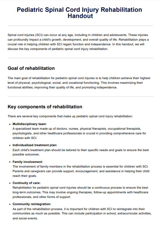
Depending on the diagnosis, lifestyle modifications may be suggested to manage symptoms or prevent further progression. This could include recommendations for changes in diet, exercise, or other aspects of daily living that may impact visual and neurological health.
Commonly asked questions
A neuro eye exam goes beyond a regular eye exam by focusing on the intricate connection between the eyes and the neurological system. While a routine eye exam primarily assesses visual acuity and eye health, a neuro eye exam delves into aspects like cranial nerve function, depth perception, and neurological conditions affecting vision.
A neurological exam evaluates the functioning of the nervous system, including the brain, spinal cord, and peripheral nerves. In the context of a neuro eye exam, specific attention is given to cranial nerves, optic nerve function, and any signs of neurological disorders impacting vision or eye movement.
During a neuro eye exam, patients can expect a thorough assessment of visual function, including tests for depth perception, visible field defects, and cranial nerve function. The examination may involve specialized equipment such as a slit lamp biomicroscope and pupillometer. The process aims to identify neurological issues affecting the eyes and ensure a comprehensive understanding of the patient's visual health.


