Neuro Ophthalmology Exam

Explore our comprehensive neuro-ophthalmology exam guide aiding precision diagnostics, tailored treatment plans, and expert care for optimal eye and brain health.

By Emma Hainsworth on Aug 08, 2024.

tick

Fact Checked by RJ Gumban.

Use Template
AI IconToolbarShare ui

What is a neuro-ophthalmology examination?

A neuro-ophthalmology examination is a specialized medical assessment focused on the complex relationship between the nervous and visual systems. Neuro-ophthalmology is an interdisciplinary field that combines elements of both neurology and ophthalmology to diagnose and treat disorders that affect the optic nerve, the visual pathways, and the eye movement systems.

Critical components of a neuro-ophthalmological examination typically include:

  • Visual acuity testing assesses how well the patient can see at various distances. It's fundamental in detecting vision loss or changes in visual acuity.
  • Visual field testing: This test measures the full horizontal and vertical range of what a patient can see peripherally. It helps identify blind spots (scotomas), indicating optic nerve damage or brain abnormalities.
  • Color vision testing: Changes in color vision can be an early sign of optic nerve disease.
  • Pupillary reflexes: Examining how pupils react to light can provide information about the optic nerve’s health and whether there's a potential neurological disorder.
  • Ophthalmoscopy: This is an examination of the back of the eye (the retina), focusing on the optic nerve head (optic disc), which can provide clues about various neurological conditions.
  • Eye movement testing evaluates the cranial nerves and brain regions controlling eye movements. Abnormalities in eye movements can indicate problems in the brain or nerves.
  • Stereopsis (depth perception) testing: This test evaluates the ability to perceive the world in three dimensions and the distance of an object.
  • External examination of the eyes and eyelids: This involves evaluating the external structures of the eye and surrounding tissues.

A neuro-ophthalmology examination aims to detect abnormalities that may point to neurological conditions such as multiple sclerosis, brain tumors, stroke, myasthenia gravis, or other systemic diseases that can affect vision and eye movements. The examination can be complex and often involves a detailed assessment of the history of symptoms and various specialized tests. It’s typically performed by a neuro-ophthalmologist, a medical doctor specializing in neurology and ophthalmology.

When would you conduct a neuro-ophthalmological assessment?

A neuro-ophthalmological assessment is conducted for several vital reasons, primarily to diagnose and manage conditions affecting the intricate relationship between the nervous and visual systems. Here are key reasons for conducting such an assessment:

  • To diagnose disorders involving the optic nerve: Conditions like optic neuritis, optic neuropathy, and ischemic optic neuropathy can directly impact the optic nerves, leading to vision loss or other visual abnormalities. These conditions require specialized attention that a neuro-ophthalmological assessment provides.
  • To evaluate issues with eye movements: Problems with eye movements can be due to disorders affecting the cranial nerves, brainstem, or neuro-muscular junctions, such as in cases of myasthenia gravis, cranial nerve palsies, or brain tumors. A neuro-ophthalmological exam helps in diagnosing these issues.
  • To investigate causes of visual field loss: Loss of peripheral vision or the presence of blind spots (scotomas) can indicate neurological issues like stroke, brain tumors, or glaucoma. Neuro-ophthalmological assessments are crucial in mapping visual fields and identifying the underlying causes.
  • To diagnose and manage double vision (diplopia): Double vision can be a symptom of various conditions, including those affecting the cranial nerves (like the third, fourth, or sixth nerve palsies), thyroid eye disease, or brain lesions. A neuro-ophthalmologist can determine the cause and recommend appropriate treatment.
  • In the presence of neurological conditions with ocular manifestations: Neurological diseases such as multiple sclerosis, Parkinson's disease, and others can have ocular manifestations. A neuro-ophthalmological assessment helps in identifying and managing these visual symptoms.
  • For evaluating pupillary abnormalities: Abnormalities in pupil size, shape, or reactivity can be signs of severe conditions like Horner's syndrome or third nerve palsy. These require a detailed neuro-ophthalmological evaluation.
  • To assess visual symptoms in systemic diseases: Certain systemic diseases like lupus or sarcoidosis can have ocular manifestations. Neuro-ophthalmological assessments help evaluate the extent and impact of these diseases on the visual system.
  • Following head trauma or neurosurgery: Patients who have suffered head trauma or have undergone neurosurgery often require a neuro-ophthalmological evaluation to assess any impact on their vision or eye movement functions.
  • To differentiate between ophthalmological and neurological causes of visual problems: When visual symptoms are present, it can be challenging to determine if they are due to primary eye diseases or neurological disorders. A neuro-ophthalmological assessment can help in making this distinction.
  • For complex cases requiring a multidisciplinary approach: Some cases may present with symptoms that overlap between neurology and ophthalmology, requiring a comprehensive approach for accurate diagnosis and treatment.

In summary, neuro-ophthalmological assessments are crucial for various conditions affecting vision and eye movements, offering specialized insights beyond the scope of standard ophthalmic or neurological evaluations.

Equipment required for a neuro-ophthalmological assessment

A neuro-ophthalmological assessment requires specialized equipment to thoroughly evaluate the complex interplay between the nervous and visual systems. Here is a list of crucial equipment typically used:

  • A Snellen chart or similar visual acuity test tool is used to measure visual acuity. These charts contain letters or symbols in various sizes and are essential for determining the sharpness or clarity of the patient's vision.
  • Perimetry machine (visual field tester): This device assesses the patient's visual field. It helps detect blind spots (scotomas), peripheral vision loss, or other visual field defects.
  • Color vision test charts or books (like Ishihara plates): These are used for evaluating color vision and identifying color vision deficiencies.
  • Pupillometer or bright light source for pupillary reaction tests: A pupillometer is a device used to measure pupil size and reactivity. Alternatively, a bright light source can also be used to assess pupillary light reflexes manually.
  • Ophthalmoscope or fundus camera: An ophthalmoscope is used to visualize the interior structures of the eye, including the retina and optic nerve. A fundus camera might capture detailed images of the retina and optic nerve.
  • Slit lamp biomicroscope: This equipment allows detailed examination of the anterior and posterior segments of the eye. It's essential for assessing the health of the eye's external and internal structures.
  • Prisms and occluders for eye movement testing: Prisms can be used to measure and diagnose deviations in eye alignment and occluders to cover one eye at a time during certain examination parts.
  • Neurological examination tools include reflex hammers and tuning forks, used for general neurological assessments that might be relevant in a neuro-ophthalmological evaluation.
  • Stereopsis testing tools: Tests like the Titmus Fly or Randot Stereotest assess depth perception.
  • Gonioscope: Used for examining the anterior chamber angle of the eye, which can be crucial in certain glaucoma cases or other conditions affecting intraocular pressure.
  • Optical coherence tomography (OCT) machine: An advanced imaging device that provides high-resolution images of the retina, optic nerve, and nerve fiber layer, helpful in diagnosing and monitoring glaucomatous damage and other retinal conditions.
  • Automated refractometer: To measure the refractive error of the eyes.
  • Tonometer: For measuring intraocular pressure, which is crucial in assessing for glaucoma and other conditions that may cause or result from increased pressure inside the eye.

This equipment comprehensively evaluates various aspects of vision and ocular health, which are critical in diagnosing and managing neuro-ophthalmological conditions.

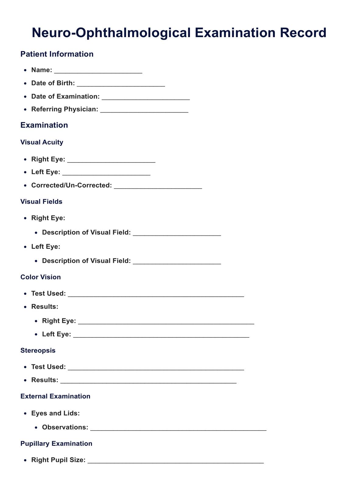
How to use this exam template

Preparation:

  • Print or digitize the template: Have the template ready in a convenient format. Depending on your record-keeping system, it could be a printed copy or a digital version.
  • Familiarize yourself with each section: Understand what each section is for and the type of information it's designed to capture.

Patient information section:

  • Fill out basic details: At the time of the examination, fill in the patient's name, date of birth, date of examination, and the referring physician’s name. This information is crucial for record identification and management.

Visual acuity testing:

  • Conduct the test: Use a Snellen chart or similar tool to test the patient's visual acuity.
  • Record the results: Enter the acuity measurements for both eyes, noting if the vision was tested with or without correction (glasses or contact lenses).

Visual field testing:

  • Perform the test: This could be done using automated perimetry or manual methods like the Goldmann field test.
  • Document findings: Record any abnormalities or deficits in the patient's peripheral vision for each eye.

Color vision testing:

  • Administer the test: Typically done using Ishihara plates or similar tests.
  • Log the outcome: Note down the results, specifying if there were any difficulties in color differentiation.

Pupillary reflexes:

  • Examine the pupils: Check for pupil size, shape, and reaction to light and accommodation.
  • Record observations: Document the findings, including any asymmetry or abnormal responses.

Ophthalmoscopic examination:

  • Inspect the optic disc and retina: Examine the back of the eye using an ophthalmoscope.
  • Note any abnormalities: Detail any optic disc, retina, or blood vessel irregularities.

Eye movement testing:

  • Assess eye movements: Check for smoothness, coordination, and involuntary or misalignments.
  • Fill in the results: Describe any deviations from regular eye movements.

Stereopsis testing:

  • Perform depth perception tests: Use tests like the Titmus Fly or Randot Stereotest.
  • Enter the results: Indicate the level of depth perception and any anomalies.

External examination:

  • Inspect external eye structures: Look at the eyelids, conjunctiva, cornea, and overall eye appearance.
  • Document findings: Any abnormalities or concerns should be recorded.

Overall assessment and notes:

  • Diagnosis and observations: Document any diagnoses or notable observations based on the test results.
  • Recommendations: Include any recommendations for treatment, follow-up, or referrals to other specialists.

Signatures:

  • Ensure completion: Sign the document once the examination is complete and all sections are filled out.
  • Obtain patient signature: If necessary, have the patient or their guardian sign, especially if consent is required for the documented findings or further procedures.

You can leverage the Myasthenia Gravis Evaluation Template to enhance your practice and improve client outcomes. This template provides a structured approach to assessing and monitoring symptoms of myasthenia gravis.

Interpreting the results

Interpreting the results of a neuro-ophthalmological assessment involves a detailed analysis of various diagnostic tests and clinical findings. The interpretation requires an understanding of the complex interactions between the nervous system and the visual system. Here’s how different aspects of the assessment are typically interpreted:

Visual acuity testing:

  • Normal results: Clear, sharp vision usually indicated by 20/20 acuity.
  • Abnormal results: Decreased acuity can indicate various issues, from refractive errors to diseases affecting the optic nerve or central nervous systems, such as optic neuritis or macular degeneration.

Visual field testing:

  • Normal results: An entire field of vision without significant blind spots.
  • Abnormal results: Deficits in the visual field can indicate optic nerve damage, retinal diseases, or neurological conditions affecting the visual pathways in the brain, such as stroke or brain tumors.

Color vision testing:

  • Normal results: Accurate color discrimination.
  • Abnormal results: Difficulties in color perception may signal optic nerve diseases or neurologic disorders affecting color processing areas in the brain.

Pupillary responses:

  • Normal results: Pupils that are equal, round, and reactive to light and accommodation.
  • Abnormal results: Anisocoria (unequal pupil sizes), poor light response, or a relative afferent pupillary defect (RAPD) can suggest optic nerve disorders or other neurologic conditions.

Ophthalmoscopy (examination of the optic disc and retina):

  • Normal results: A healthy optic disc and retina without swelling, hemorrhages, or abnormal vessels.
  • Abnormal results: Findings like papilledema (optic disc swelling) can indicate increased intracranial pressure; optic atrophy might suggest chronic optic nerve damage.

Eye movement examination:

  • Normal results: Full and smooth eye movements in all directions.
  • Abnormal results: Restricted movement, nystagmus, or double vision can point to problems with the cranial nerves, brainstem, or cerebellum.

Optical coherence tomography (OCT):

  • Normal results: A standard retinal nerve fiber layer and macular thickness.
  • Abnormal results: Thinning of the nerve fiber layer or changes in the macula can indicate glaucoma, optic neuropathies, or macular diseases.

Assessment of eyelids and external eye structures:

  • Normal results: Healthy appearance of the eyelids and surrounding tissues.
  • Abnormal results: Ptosis (drooping eyelid), lid retraction, or eyelid swelling can be associated with cranial nerve palsies, thyroid eye disease, orbital diseases.

The interpretation of these results should be contextualized within the patient’s overall health, symptoms, and medical history. Abnormal findings often warrant further investigation, potentially including additional specialized tests, consultations with other medical specialists, or ongoing monitoring. The ultimate goal of interpreting these results is to understand the patient's condition comprehensively, leading to an accurate diagnosis and effective treatment plan.

Next steps

Steps involve a series of actions to ensure optimal patient care and management. These steps are crucial for addressing the identified issues and planning the appropriate course of treatment or further investigation:

  • Discuss results with the patient: Explain the examination findings to the patient. Ensure they understand their condition, the implications of the test results, and the need for further testing or treatment.
  • Develop a treatment plan: Based on the examination results, devise a treatment plan. This may include medical therapy, lifestyle modifications, referrals to specialists, or considering surgical options if applicable.
  • Prescribe medications or therapies: If the diagnosis warrants, prescribe the appropriate medications or therapies. For instance, in cases of optic neuritis, steroid therapy might be initiated.
  • Referrals to specialists: For conditions that require specialized care, such as thyroid eye disease, myasthenia gravis, or advanced glaucoma, refer the patient to appropriate specialists like endocrinologists, neurologists, or glaucoma specialists.
  • Order additional diagnostic tests: If the initial examination suggests conditions like optic neuropathy or macular degeneration, additional tests like optical coherence tomography (OCT), MRI, or fluorescein angiography might be necessary for a more definitive diagnosis.
  • Schedule follow-up appointments: Arrange for follow-up visits to monitor the patient's condition, evaluate the effectiveness of treatment, and make any necessary adjustments to the care plan.
  • Patient education and support: Provide educational resources to help patients understand their condition. This might include information about managing symptoms, potential lifestyle adjustments, and understanding the long-term outlook of their condition.
  • Coordinate with primary care providers: Communicate with the patient's primary care provider to ensure a comprehensive approach to their overall health, particularly if the patient has systemic conditions affecting their vision.
  • Documentation and record keeping: Accurately document all findings, treatment plans, patient interactions, and referrals. This is crucial for ongoing patient care and for any future medical needs.
  • Continued professional development: Stay updated with the latest advancements and research in neuro-ophthalmology to enhance patient care continually. This may include attending conferences, participating in clinical trials, and reading current literature.
  • Patient monitoring and long-term management: Some neuro-ophthalmological conditions require long-term monitoring and management. Set up a long-term care plan with regular check-ups and evaluations as needed.

These steps ensure the patient receives comprehensive care tailored to their neuro-ophthalmological needs, leading to the best possible outcomes.

What does a neuro-ophthalmological assessment involve for patients with complaints of visual loss, particularly in distance vision and central vision?

Commonly asked questions

What does a neuro-ophthalmological assessment involve for patients with complaints of visual loss, particularly in distance vision and central vision?

A neuro-ophthalmological assessment for visual loss includes evaluating the best corrected visual acuity for distance and central vision. Tests like fundus examination and optical coherence tomography (OCT) assess the health of the optic nerves and macula. The assessment may include pupil testing, confrontation visual fields, and a detailed review of any refractive error. The neuro-ophthalmologist aims to diagnose macular degeneration, optic neuritis, or central serous retinopathy.

How do neuro-ophthalmologists approach differential diagnosis in patients with peripheral vision loss and other visual complaints?

Neuroophthalmologists use a combination of patient history, clinical examination, and diagnostic tests to perform differential diagnosis. They assess peripheral vision loss through visual field testing and examine the optic disc with a slit lamp or OCT. Conditions like glaucoma, stroke, and optic nerve diseases are considered. Pupil testing and examination of eyelid abnormalities may also provide crucial diagnostic information.

What specific tests are conducted in cases of suspected optic nerve disorders like optic neuritis or dominant optic atrophy?

Neuro-ophthalmologists conduct a detailed eye examination for suspected optic nerve disorders, including a fundus examination to visualize the optic disc. OCT is used to assess the optic nerve fiber layer, and visual field tests check for characteristic patterns of visual loss. Additional tests might include an MRI to visualize the optic nerves and assess for demyelinating diseases in cases like optic neuritis.

Join 10,000+ teams using Carepatron to be more productive

One app for all your healthcare work