Eye Physical Examination
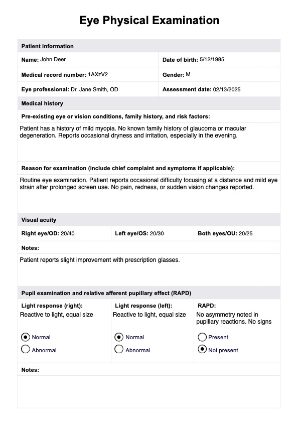
An eye physical exam assesses vision and eye health and detects any abnormalities or diseases. Download Carepatron's free eye exam PDF here.

What is an Eye Physical Examination?
An eye exam, also known as an ophthalmic or optometric examination, is a comprehensive assessment of the visual health and function of the eyes. It involves a series of tests and evaluations performed by an eye care professional, such as an optometrist or ophthalmologist, to determine the overall condition of the eyes and identify any potential issues.
Various aspects of vision and eye health are scrutinized during an eye exam. The process typically includes measuring visual acuity, assessing refractive error (nearsightedness, farsightedness, or astigmatism), and examining the eye's internal and external structures, such as the cornea, lens, pupil, and iris eyelid of the left and right eyes.
An eye exam is not limited to individuals with vision problems. Those with normal vision should undergo periodic eye exams to maintain eye health. As many cases of vision loss and eye conditions (including certain glaucoma and macular degeneration types) are preventable, regular eye exams are key for early detection of potential problems.
A comprehensive physical examination of the eyes involves a series of assessments to evaluate various aspects of visual health and detect potential issues that may affect the eyes or vision. Here are some common components:
Visual acuity testing
Visual acuity testing measures the sharpness and clarity of vision, typically using a Snellen chart (where letters or symbols are read from a standardized distance). This assessment helps identify refractive errors such as myopia (nearsightedness), hyperopia (farsightedness), or astigmatism.
Pupils
This test involves assessing the size, equality, and response to light of the pupils. Relative afferent pupillary (RAPD) may be checked by shining a light into one pupil at a time and observing the constriction response in both pupils. This test provides valuable information about optic nerve function and potential neurological issues.
Extraocular motility and alignment
Evaluating extraocular motility and alignment assesses the coordination and movement of the eyes. It helps identify conditions such as strabismus (misalignment) and ensures proper eye teaming for binocular vision.
Intraocular pressure
This test is performed using a tonometer and is crucial for detecting glaucoma. Increased intraocular pressure can damage the optic nerve over time.
Confrontation visual fields
This test checks the patient's ability to see objects in their peripheral vision without directly looking at them. This test can reveal defects in the visual field, which may be indicative of various eye or neurological problems.
External examination
The external examination involves inspecting the eyelids, lashes, and surrounding tissues for abnormalities. Any signs of inflammation, infection, or other issues are carefully noted during this part of the exam.
Slit-lamp examination
The slit-lamp examination provides a detailed view of the anterior and posterior segments of the eye. It helps identify abnormalities in the cornea, lens, and other structures that may contribute to eye problems.
Fundoscopic examination
This technique typically involves stabilizing the patient's shoulder and using an ophthalmoscope to examine the interior of the eye, including the optic nerve, retina, and blood vessels. This is crucial for detecting conditions like diabetic retinopathy and macular degeneration.
Eye Physical Examination Template
Eye Physical Examination Example
How to use the Eye Physical Examination template
This complete eye exam template is a practical tool designed to guide individuals through a structured, comprehensive eye examination process. Here are some steps when using the complete eye exam template:
Step 1: Access the template
To access the template, click the "Use template" button on this page to open the template in the Carepatron app. Alternatively, click the "Download" button to access a ready-to-go PDF version.
Step 2: Medical history
Record the reason for the examination (e.g. routine check-up). Record any specific symptoms (e.g. blurry or double vision, eye pain), family history, or risk factors of eye disease (e.g. high blood pressure).
Step 3: Visual acuity assessment
Use an eye chart or a visual acuity app to measure the clarity of vision. Record the most minor line of text that can be read accurately.
Step 4: Pupil Examination and RAPD
Record dilated pupil size, equality, and reaction to light reflection in both eyes. Assess for any signs of a defect, which may indicate optic nerve dysfunction.
Step 5: Evaluate ocular mobility
Assess eye movement by checking for smooth pursuit, saccades, and convergence (e.g. using a 'swinging flashlight' test). Record any irregularities in the movement of the eyes that may warrant further investigation into potential eye or vision problems.
Step 6: Visual field test
Conduct a confrontation visual field test to evaluate peripheral vision. This step helps identify abnormalities or blind spots in the visual field, providing insights into potential eye conditions.
Step 7: Interior and external eye examination
Inspect the external structures of the eye, including the eyelids, lashes, and surrounding tissues. Look for signs of inflammation, infection, or other eye health issues. If available, use a slit lamp or a pen bright light to examine the anterior chamber of the eye, including the cornea, lens, and other structures for abnormalities. If a fundoscopic examination of the eye's interior is performed, record the health of the optic nerve, retina, and blood vessels.
Results and interpretation
Here, are some of the most common outcomes of a comprehensive optometry examination and what they signify:
Visual acuity
- Normal: the eyes can focus on objects at a standard distance. Regular monitoring is still essential to catch any changes in visual acuity over time.
- Abnormal: corrective lenses may be necessary.
Equal pupils and responsive to light
- Normal: a healthy pupillary response is indicated by equal-sized pupils, that are appropriately responsive to light.
- Abnormal: any significant differences in pupil size or a lack of reaction could indicate issues such as nerve damage or neurological conditions (e.g. optic neuritis), necessitating further investigation.
Eye movement and alignment
- Normal: normal range eye movement and alignment suggest proper coordination, enabling effective binocular vision.
- Abnormal: if irregularities, such as saccades, might indicate problems with eye muscle control or alignment issues, warranting a closer examination.
Intraocular pressure
- Normal: a normal intraocular pressure reading suggests low risk of glaucoma.
- Abnormal: increased pressure may suggest ocular hypertension or glaucoma. Low pressure indicates insufficient aqueous humour production, which can be caused by chronic inflammation or retinal detachment.
Visual field
- Normal: a full and even visual field is indicative of a healthy peripheral vision.
- Abnormal: Any deficits or blind spots detected during confrontation visual field testing may necessitate further investigation for conditions affecting the optic nerve or visual pathways.
External examination
- Normal: healthy eyelids, lashes, and surrounding tissues.
- Abnormal: Any signs of redness, swelling, or abnormalities may warrant closer inspection and evaluation for potential infections or inflammations.
Clear slit-lamp and fundoscopic examination
- Normal: a clear view during slit-lamp and fundoscopic examinations indicates the absence of abnormalities in the cornea, lens, optic nerve, retina, and blood vessels.
- Abnormal: clouding of the lens (cataracts), corneal injuries, presence of foreign objects, infections like conjunctivitis, macular degeneration, dry eye syndrome, hyphema, and retinal detachment may all be detected in a slit-lamp exam. Abnormal findings of a fundoscopic exam include swollen optic disc or cup, obscured red reflex, uveal melanoma, neovascularization and microaneuryisms.
Other eye tests
Additional tests may also be used in evaluating ocular health and visual function, depending on the patient's specific needs or the eye professional's usual practice. Here are some other types of eye tests:
Retinoscopy
Retinoscopy is a technique used to determine refractive error. By shining a light into the eye and observing the reflection of the retina, eye care professionals can assess the need for corrective lenses.
Color vision testing
Color vision testing evaluates the ability to perceive and distinguish various colors. Commonly conducted using Ishihara plates, this test is particularly crucial for occupations where accurate color perception is essential. Difficulty discerning colors can affect one's ability to perform certain tasks or lead to blurry vision.
Refraction
Refraction is used to determine the best prescription for corrective lenses. By assessing how light bends as it enters the eyes, eye care professionals can precisely prescribe glasses or contact lenses.
Corneal topography
Corneal topography is a specialized test mapping the curvature of the cornea's surface. It provides detailed information about the cornea's shape and helps diagnose conditions affecting vision, such as astigmatism. This test particularly benefits those considering laser eye surgery or contact lens fitting.
Commonly asked questions
A physical exam of the eyes involves a series of assessments to evaluate visual acuity, eye movement, pupil reactions, and the health of ocular structures. Eye care professionals follow a comprehensive process to ensure all crucial aspects of eye health and function are assessed.
A routine eye exam typically includes visual acuity testing, pupil assessment, evaluation of extraocular movements and extraocular muscles, confrontation visual field testing, external examination, and various specialized tests based on individual needs. If the patient has a family history or particular risk factors for a particular eye condition, the examiner may perform additional testing.
The appropriate frequency of eye exams depends on the patient's individual health, family history and risk factors. Generally, patients should aim to present for eye testing approximately every 2 years, but more frequent exams are advisable if specific symptoms such as eye pain or vision loss occur.