Doppler Ultrasound
Understand the Doppler Ultrasound Test, a non-invasive procedure that estimates blood flow through your vessels using high-frequency sound waves. Learn more today.
�?�
What is a Doppler Ultrasound Test?
A Doppler ultrasound is a specialized, non-invasive medical procedure that utilizes high-frequency sound waves to assess blood movement through your vessels. The process involves a handheld device known as a transducer, gently moving across the body's area under examination. As it moves, it emits and picks up sound waves. A computer system then processes these waves, resulting in images and data that provide a detailed account of blood flow.
This diagnostic tool is crucial in detecting and assessing conditions that affect blood flow, such as venous insufficiency, arterial blockages, and blood clots. Its significance lies in its ability to detect potential blood circulation issues efficiently and painlessly, making it invaluable in various medical fields. For instance, it's commonly used during pregnancy to monitor fetal health, providing insights into the unborn child's well-being. Neurology is employed to evaluate stroke risk by analyzing the blood flow in the brain, thus aiding in early detection and prevention.
The Doppler ultrasound test provides information that helps healthcare professionals make informed decisions about patient care. It offers a safe, effective, and quick method to gain vital insights into the body's circulatory system. Its non-invasive nature also poses minimal risks, making it a preferred choice for clinicians and patients.
Doppler Ultrasound Template
Doppler Ultrasound Example
How Does it Work?
By bouncing high-frequency sound waves off circulating red blood cells, the Doppler ultrasound test can detect changes in the frequency of the reflected waves depending on the speed and direction of the blood flow. This change in frequency is then calculated by the machine and displayed visually, providing a detailed map of the blood flow velocity.
Here's a step-by-step breakdown of how this process unfolds:
Preparing for the Test
Your Doppler ultrasound test begins with preparation. You'll typically be asked to wear a hospital gown for easy access to the area being examined. After changing, you'll be instructed to lie on an examination table in a position that allows the sonographer to perform the scan easily.
Applying the Gel
Next, the sonographer will apply a special water-based gel to the skin over the examined area. This gel serves a crucial purpose - it aids in penetrating the sound waves into your body, ensuring clear, high-quality images.
Scanning with the Transducer
The sonographer will then move the transducer - a handheld device that emits and receives sound waves - across the gelled area. As the transducer moves, sound waves bounce off the circulating red blood cells and return to the device.
Interpreting the Results
The returned sound waves are transformed into a visual representation of blood flow on a computer screen. A trained professional can then interpret these images for diagnosis or monitoring purposes.
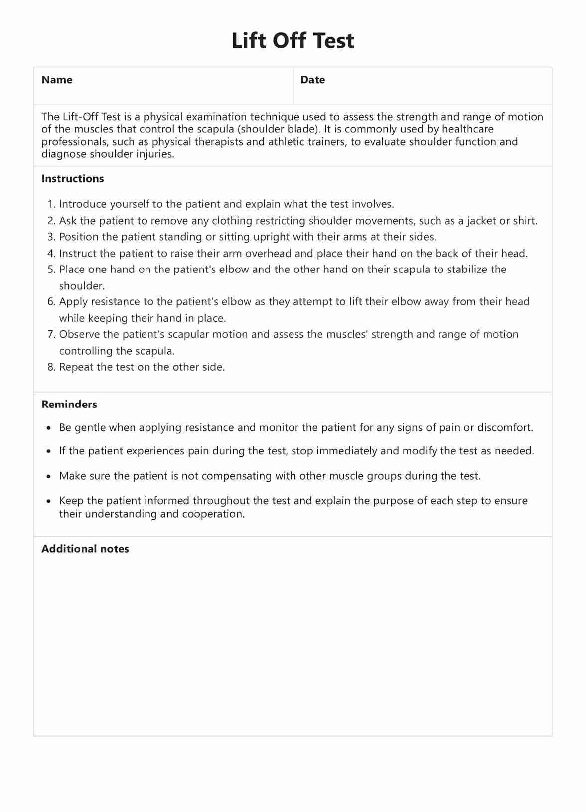
We have a printable Doppler Ultrasound Test, offering a comprehensive and visual insight into your blood flow, aiding healthcare professionals in making informed medical decisions.
When would you use this test?
The Doppler ultrasound test is a versatile medical tool employed across various fields of medicine for different purposes. Its primary function is to provide a detailed look at blood flow within the body's vessels, making it an invaluable resource in diagnosing and monitoring conditions that affect these vessels.
One of the most common uses of this test is in diagnosing vascular conditions. It can detect abnormalities such as blood clots, blockages, or insufficiencies in veins and arteries. This makes it an essential tool for cardiologists and vascular surgeons who regularly deal with these issues.
In obstetrics, the Doppler ultrasound test is pivotal in prenatal care. It can monitor the fetus's health by assessing blood flow in the umbilical cord, placenta, and other fetal vessels. This helps healthcare providers ensure the well-being of both the mother and the baby during pregnancy.
Neurology also benefits from the Doppler ultrasound test, specifically through a variation known as transcranial Doppler ultrasound. This test evaluates blood flow in the brain, assisting in assessing stroke risk. Identifying potential issues early aids in preventive care and enhances patient outcomes.
Additionally, the Doppler ultrasound test can be used post-treatment to monitor the success of interventions for vascular conditions. It can track improvements in blood flow after procedures like angioplasty or bypass surgery, providing doctors with crucial feedback.
The Doppler Ultrasound Test is a powerful diagnostic tool that helps healthcare professionals make informed decisions about patient care. Its versatility and non-invasive nature make it an ideal choice across various medical disciplines.
What do the results mean?
The results of a Doppler ultrasound test offer a comprehensive look at the body's blood flow, providing invaluable information for diagnosing and monitoring various health conditions. While the specifics can vary depending on the individual and the area being examined, here are some common interpretations:
Normal results from a Doppler ultrasound test typically indicate that blood flows freely through the body's vessels without any significant obstructions or abnormalities. This is often denoted by smooth, regular waveforms on the test results, signifying healthy blood flow. On the other hand, abnormal results can suggest a range of potential issues. For instance, reduced blood flow might indicate a blockage or narrowing in a blood vessel due to conditions such as arteriosclerosis or deep vein thrombosis.
Conversely, increased blood flow could signal inflammation in the area or the presence of an arteriovenous malformation (an abnormal connection between arteries and veins). In pregnancy, abnormal blood flow in the umbilical cord may indicate potential complications like intrauterine growth restriction.
It's important to note that the results of a Doppler ultrasound test aren't interpreted in isolation. To make a comprehensive assessment, a healthcare professional will consider these findings in conjunction with your symptoms, medical history, and overall health.
Remember, a Doppler ultrasound test is a non-invasive, risk-free procedure that can provide valuable insights into your vascular health. Our Free Doppler Ultrasound Test can help you understand this process better and prepare you for what to expect.
Research & Evidence
The Doppler ultrasound has a well-established history and robust body of research supporting its effectiveness in various medical scenarios. Pioneered in the 1950s, it has evolved over the decades into an indispensable tool for diagnosing and monitoring various vascular conditions.
Early pioneers, like Callagan, laid the groundwork by conducting seminal research using Doppler ultrasound to evaluate patients with peripheral vascular disease. Subsequent studies have built on this foundational work, confirming the accuracy and utility of Doppler ultrasound across diverse clinical settings.
For instance, research published in the journal "Rheumatology" underscored the importance of Doppler ultrasound in predicting late pregnancy outcomes in patients with systemic lupus erythematosus. Other studies, such as one published in "Annals of Biomedical Engineering," highlighted the precision of Doppler ultrasound in assessing aortic wave reflection in children and adolescents with a history of heart disease.
Moreover, Doppler ultrasound has proven instrumental in obstetrics and gynecology, diagnosing conditions like placenta previa accreta and monitoring fetal health. It's also been effectively used in neurology for evaluating cerebrovascular health.
The rich history of Doppler ultrasound and the extensive body of research supporting its use underscore its value in modern medicine. Its continued refinement and adaptation to new clinical scenarios speak to its enduring relevance and utility.
References
- Aaslid, R., Markwalder, T. M., & Nornes, H. (1982). Noninvasive transcranial Doppler ultrasound recording of flow velocity in basal cerebral arteries. Journal of neurosurgery, 57(6), 769-774. Retrieved from https://link.springer.com/content/pdf/10.1007/978-94-009-8204-8.pdf.
- Abramowicz, J. S., & Sheiner, E. (2008). In utero imaging of the placenta: importance for diseases of pregnancy. Placenta, 29, S14-S22. Retrieved from https://www.medultrason.ro/medultrason/index.php/medultrason/article/view/3845.
- Baker, L. E. (1957). The history of ultrasound. Ultrasound in Medicine & Biology, 33(6), 809??839. Retrieved from https://www.sciencedirect.com/science/article/pii/S003961090570308X.
- Costedoat-Chalumeau, N., Amoura, Z., Lupoglazoff, J. M., Huong, D. L., Denjoy, I., Vauthier, D., ... & Piette, J. C. (2006). Outcome of pregnancies in patients with anti-SSA/Ro antibodies: a study of 165 pregnancies, with special focus on electrocardiographic variations in the children and comparison with a control group. Arthritis & Rheumatism: Official Journal of the American College of Rheumatology, 54(11), 3305-3311. Retrieved from https://onlinelibrary.wiley.com/doi/abs/10.1002/art.22199.
- Kheyfets, V. O., Schafer, M., Podgorski, C. A., Schroeder, J. D., Browning, J., Hertzberg, J., ... & Hunter, K. S. (2017). Patient-specific computational modeling of blood flow in the pulmonary arterial circulation. Computer methods in biomechanics and biomedical engineering, 20(9), 942-954. Retrieved from https://link.springer.com/article/10.1007/s10439-023-03339-2.
- Ofili-Yebovi, D., Ben-Nagi, J., Sawyer, E., Yazbek, J., Lee, C., Gonzalez, J., ... & Jurkovic, D. (2008). Deficient lower-segment Cesarean section scars: prevalence and risk factors. Ultrasound in Obstetrics and Gynecology: The Official Journal of the International Society of Ultrasound in Obstetrics and Gynecology, 31(1), 72-77. Retrieved from https://obgyn.onlinelibrary.wiley.com/doi/abs/10.1046/j.1469-0705.2000.00018.x.
- Satomura, S. (1986). History of pulse and echo Doppler ultrasound in Japan. In: White DN. (ed) Blood Flow Measurement. Palgrave, London. Retrieved from https://link.springer.com/chapter/10.1007/978-94-009-4988-1_2.
- Strandness, D. E., Jr. (1998). A brief history of Doppler ultrasound in the diagnosis of peripheral vascular disease. Journal of Vascular Surgery, 27(3), 602??607. Retrieved from https://www.sciencedirect.com/science/article/pii/S0301562997002640.
Commonly asked questions
Various healthcare professionals may request a Doppler ultrasound test, including cardiologists, vascular surgeons, obstetricians, and neurologists.
Doppler ultrasound tests are used when there is a need to assess the blood flow in the body's vessels. This may be for diagnosing conditions, monitoring treatment effectiveness, or evaluating fetal health during pregnancy.
Doppler ultrasound tests are used by applying a transducer to the skin over the area to be examined. The transducer emits and receives sound waves, which are processed into images and data about blood flow.
The length of a Doppler ultrasound test can vary depending on the area being examined, but generally, the procedure takes between 30 minutes to an hour.