Chest Pain Workup
Learn about the comprehensive workup for chest pain, including diagnostic tests and evaluations. Download a free PDF example for reference.
What is a Chest Pain Workup?
Chest pain workup is a comprehensive medical assessment designed to identify the underlying causes of chest discomfort or pain. It serves as a structured process to evaluate and diagnose the potential reasons behind chest pain, a symptom that can be associated with various medical conditions, ranging from minor issues to serious cardiac events.
During a chest pain workup, healthcare professionals employ a combination of medical history analysis, physical examination, and diagnostic tests to pinpoint the root cause of the pain. This process is crucial in distinguishing between benign causes, such as muscle strain or indigestion, and more serious conditions like heart-related issues.
The workup typically begins with a detailed discussion of the patient's medical history, including any previous heart conditions, lifestyle factors, and the specifics of the chest pain experienced. This information helps in narrowing down potential causes and guiding the subsequent steps of the assessment.
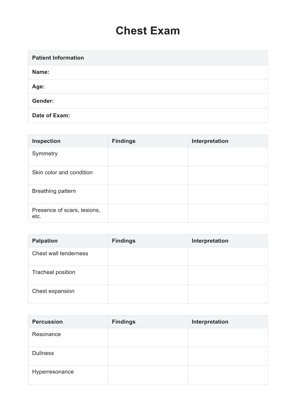
A thorough physical examination follows, where healthcare providers may assess vital signs, listen to the heart and lungs, and check for any signs of distress or abnormalities. Based on these initial assessments, healthcare professionals then determine the need for additional diagnostic tests, which may include electrocardiograms (ECGs or EKGs), blood tests, chest X-rays, and more advanced imaging studies.
Chest Pain Workup Template
Chest Pain Workup Example
The importance of documentation following a workup
In healthcare, meticulous documentation following a chest pain workup holds paramount significance. This process involves systematically recording the findings, assessments, and diagnostic outcomes obtained during the workup, creating a comprehensive record that serves multiple crucial purposes.
First and foremost, accurate documentation is instrumental in ensuring a seamless continuum of care for individuals who have undergone a chest pain workup.
For those presenting with severe chest pain, documentation becomes a critical tool in tracking the progression of their condition and aids in the identification of potential patterns or recurrent issues. This documentation not only enhances communication among healthcare providers but also facilitates a quicker response to any subsequent occurrences such as:
- Acute coronary syndrome
- Acute chest pain
- Chest wall pain
- Acute myocardial infarction.
Moreover, detailed documentation is essential for medico-legal reasons. It records the healthcare professional's diligence and adherence to standards of care during the chest pain workup. This documentation can be crucial in defending decisions made during the workup process and ensuring transparency in the provision of medical care.
In cases of acute chest pain, the documentation becomes a valuable resource for future reference. It aids in evaluating the efficacy of interventions employed during the workup and provides a baseline for monitoring the patient's progress.
Additionally, thorough documentation aids in identifying potential risk factors and establishing preventive measures, contributing to the overall management of cardiovascular health.
How to perform a chest pain assessment
Performing a chest pain assessment is a crucial skill for both healthcare professionals and individuals monitoring their own well-being. Understanding the steps involved in a thorough assessment can aid in identifying the potential causes of chest pain, including acute coronary syndrome, chest wall pain, acute myocardial infarction, and other cardiac diseases. Below are the key steps to guide you through a comprehensive chest pain assessment:
1. Patient history
Begin by obtaining a detailed patient history. Inquire about the nature and duration of the chest pain, associated symptoms, and any relevant medical history, including prior acute coronary syndrome or cardiac disease occurrences.
2. Characterization of pain
Evaluate the characteristics of the chest pain. Ask the individual to describe the pain's intensity, quality, location, and any factors exacerbating or alleviating it. Distinguish between sharp, stabbing, or pressure-like sensations.
3. Risk factors assessment
Identify potential risk factors for cardiovascular issues. Assess lifestyle factors such as smoking, diet, and exercise. Evaluate family history and the presence of conditions like diabetes or hypertension.
4. Physical examination
Perform a focused physical examination, including vital signs, heart and lung sounds, and an assessment of the chest wall. Look for signs of distress, such as sweating or pallor, which may indicate acute myocardial infarction.
5. Electrocardiogram (ECG or EKG)
Conduct an electrocardiogram to assess the electrical activity of the heart. This step is crucial in identifying abnormalities that may suggest acute coronary syndrome or cardiac issues.
6. Laboratory tests
Order relevant laboratory tests, such as cardiac enzymes and blood tests, to further evaluate the possibility of acute myocardial infarction or other cardiac conditions.
7. Imaging studies
Depending on the findings, consider imaging studies such as chest X-rays or CT scans to visualize the structures of the chest and rule out non-cardiac causes of chest pain.
8. Consultation with specialists
If necessary, refer the individual to a cardiologist or specialist for further evaluation and management of cardiac concerns.
Further information
Chest pain, that unwelcome tightness or squeezing in your chest, has been a human ailment for as long as recorded history. However, the workup to understand and diagnose the cause of this pain is a relatively recent story, filled with a fascinating medical history and evolving advancements.
References to chest pain appear in medical texts from ancient civilizations like Egypt, Greece, and India. These early descriptions focused on symptoms and potential causes, often attributing it to heart issues, lung problems, or even divine displeasure.
During the Middle Ages, physicians like Avicenna began using physical examination techniques like palpation and percussion to gather clues about the source of chest pain. Although still rudimentary, these methods marked a shift towards objective assessment.
The 17th century witnessed significant leaps in understanding anatomy and physiology. According to Friedland (2009), William Harvey's discovery of blood circulation paved the way for William Heberden's identification of angina pectoris, a specific type of chest pain related to heart disease.
Advancements like the stethoscope (René Laennec) and percussion auscultation (Jean-Nicolas Corvisart) allowed for more detailed examination of the chest and lungs, differentiating cardiac from pulmonary causes of pain.
The 20th century saw a rapid ascent of diagnostic tools. According to Barold (2003), the electrocardiogram (ECG) by Willem Einthoven in 1903 revolutionized the diagnosis of heart problems, including those causing chest pain. X-rays, followed by computerized tomography (CT) scans, offered crucial insights into lung health and potential causes of pain.
While technology has played a crucial role, the chest pain workup is not solely about machines. A thorough medical history remains essential, examining factors like onset, duration, location, and character of the pain. Assessing risk factors like smoking, high blood pressure, and family history adds valuable context.
The chest pain based workup is a testament to the continuous evolution of medical science. From ancient observations to cutting-edge technology, it showcases the unwavering human quest to understand and alleviate suffering. As the field develops, the workup promises to become even more accurate and efficient, ensuring better outcomes for patients experiencing this age-old complaint.
Chest pain risk factors
Understanding the risk factors associated with chest pain is pivotal in conducting a thorough assessment and facilitating an accurate differential diagnosis. Recognizing these factors can aid in identifying potential causes, including chest wall pain, cardiac disease, and acute coronary syndrome. Here are some key chest pain risk factors:
- Cardiovascular risk factors: Individuals with a history of cardiac disease, hypertension, or high cholesterol levels are at an increased risk of experiencing chest pain. These factors contribute to the development of atherosclerosis, a condition that may lead to acute coronary syndrome.
- Age and gender: Age plays a significant role, with older individuals more prone to cardiac-related chest pain. Additionally, men and postmenopausal women face a higher risk compared to premenopausal women.
- Smoking and tobacco use: Smoking is a significant contributor to chest pain, increasing the risk of coronary artery disease. The harmful substances in tobacco can accelerate the progression of atherosclerosis.
- Diabetes mellitus: Individuals with diabetes are at an elevated risk of developing cardiovascular issues, contributing to the likelihood of experiencing chest pain. Diabetes is often associated with accelerated atherosclerosis.
- Psychosocial factors: Stress, anxiety, and depression can influence the perception and intensity of chest pain. Addressing these psychosocial factors is crucial in managing and understanding the overall presentation.
- Physical inactivity and poor diet: Sedentary lifestyles and unhealthy dietary habits contribute to obesity and metabolic syndrome, increasing the risk of cardiovascular diseases and, subsequently, chest pain.
- Family history: A family history of cardiac disease can predispose individuals to chest pain. Genetic factors may contribute to developing conditions such as angina or myocardial infarction.
Chest pain differential diagnosis
Chest pain differential diagnosis involves systematically considering and ruling out various potential causes of chest discomfort. This process is crucial for healthcare professionals in accurately identifying the underlying issue.
The differential diagnosis for chest pain encompasses various conditions, including cardiac, pulmonary, gastrointestinal, and musculoskeletal disorders.
Key considerations during the differential diagnosis include evaluating the chest pain's nature, duration, and associated symptoms. Conditions such as angina, myocardial infarction, pulmonary embolism, gastroesophageal reflux disease (GERD), and musculoskeletal issues must be carefully considered.
Given the diverse array of possible causes, healthcare providers employ a combination of patient history, physical examination, and diagnostic tests to narrow down the potential diagnoses. Collaboration with specialists and the use of imaging studies, electrocardiograms, and laboratory tests aid in achieving a precise and thorough chest pain differential diagnosis.
Benefits of using this workup template
Efficiency and precision are paramount when dealing with patients presenting with chest pain. Carepatron's chest pain workup template offers several benefits, streamlining the assessment process and ensuring optimal outcomes for both healthcare professionals and patients.
Here are some of the key benefits of using this workup template:
Structured approach to differential diagnosis
The template provides a systematic framework for evaluating various causes of chest pain, enabling healthcare providers to conduct a thorough and efficient differential diagnosis. This structured approach helps in distinguishing between cardiac and noncardiac chest pain.
Comprehensive documentation
Utilizing the template ensures meticulous documentation of patient history, physical examination findings, and results of diagnostic tests. This comprehensive record is invaluable for ongoing patient care, consultations with specialists, and medico-legal purposes.
Improved communication
The template facilitates improved communication among healthcare team members. Clear documentation enhances the exchange of information, especially in emergency department settings, ensuring a cohesive and well-coordinated approach to patients with chest pain.
Enhanced patient understanding
Patients with chest pain often experience anxiety and uncertainty. The template allows healthcare providers to explain the assessment process more clearly, fostering a better understanding of the differential diagnosis and the steps taken to identify the cause of their discomfort.
Optimal resource utilization
By efficiently guiding the chest pain workup, the template aids in optimal resource utilization. Healthcare professionals can prioritize tests and interventions based on the identified risk factors and clinical findings, ensuring timely and cost-effective care.
Quick triage in the emergency department settings
In emergency department scenarios, where timely decisions are critical, the template expedites the triage process for patients with chest pain. This can lead to faster interventions for those with acute conditions, improving overall patient outcomes.
References
Friedland, G. (2009). Discovery of the function of the heart and circulation of blood. https://www.ncbi.nlm.nih.gov/pmc/articles/PMC3721262/#:~:text=He%20tried%20to%20force%20blood,maintained%20the%20one%2Dway%20flow.
Barold, S. S. (2003). Willem einthoven and the birth of clinical electrocardiograhy a hundred years ago. https://pubmed.ncbi.nlm.nih.gov/12766530/
Commonly asked questions
The chest pain workup involves a systematic approach to identify the underlying cause of discomfort. It typically includes a detailed patient history, physical examination, electrocardiogram (ECG or EKG), blood tests, and imaging studies, such as chest X-rays, to ensure a comprehensive assessment.
The 3 P's of chest pain refer to Provocative factors, Palliative factors, and associated Symptoms. Understanding what provokes or alleviates chest pain, along with noting associated symptoms, assists healthcare providers in forming a clearer diagnostic picture during the assessment.
Key tests for chest pain include an electrocardiogram (ECG or EKG) to assess heart function, blood tests to check for cardiac enzymes and other indicators, and imaging studies like CT scans or chest X-rays. These tests aid in differentiating between cardiac and non-cardiac causes, guiding an accurate diagnosis and appropriate treatment.