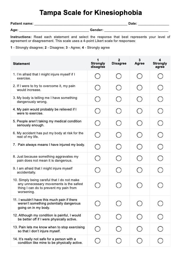
Retina Exam
Learn about retina exams: essential for detecting eye conditions like diabetic retinopathy and macular degeneration, ensuring eye health and vision safety.

Fact Checked by Ericka Pingol.
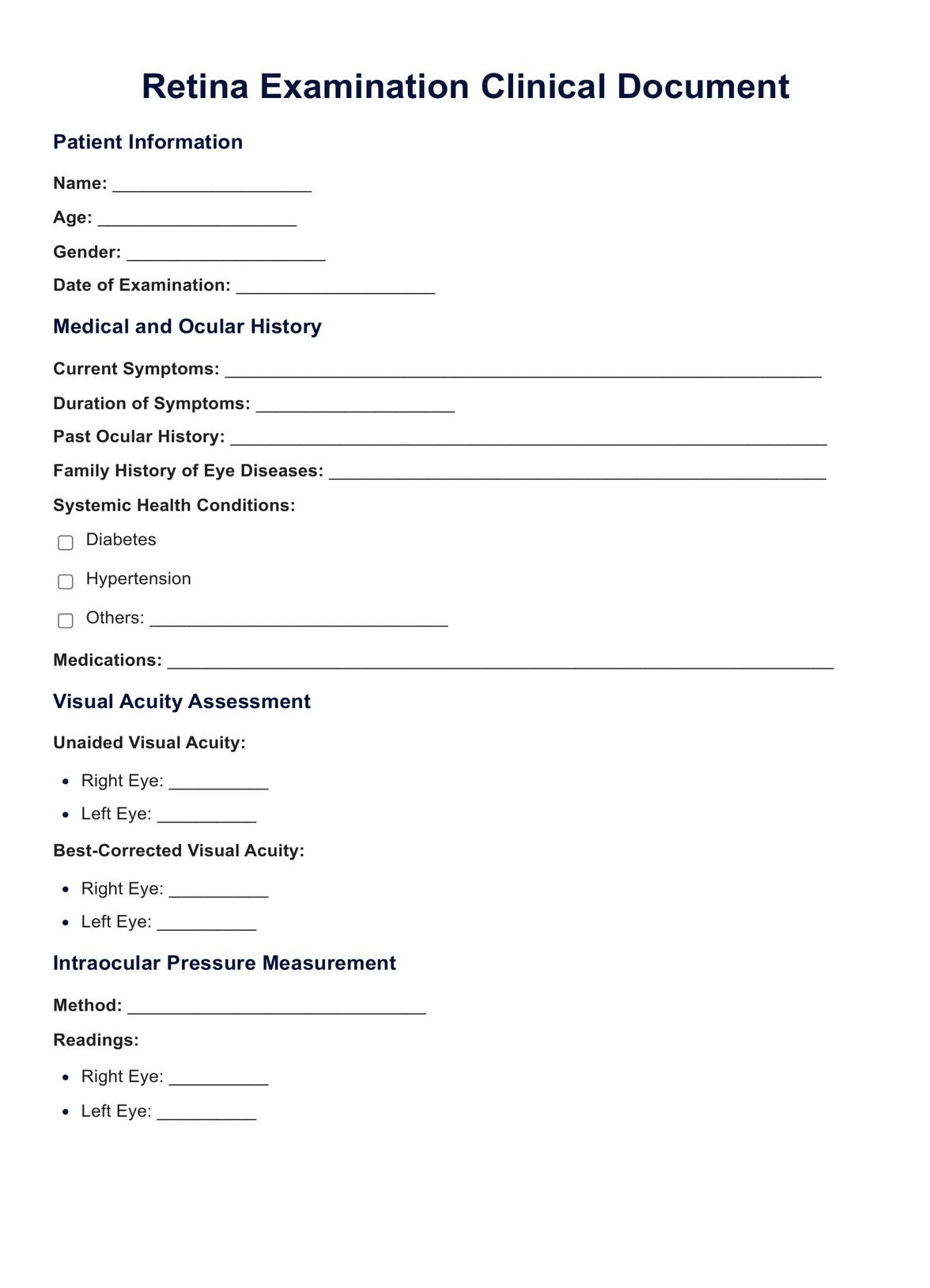


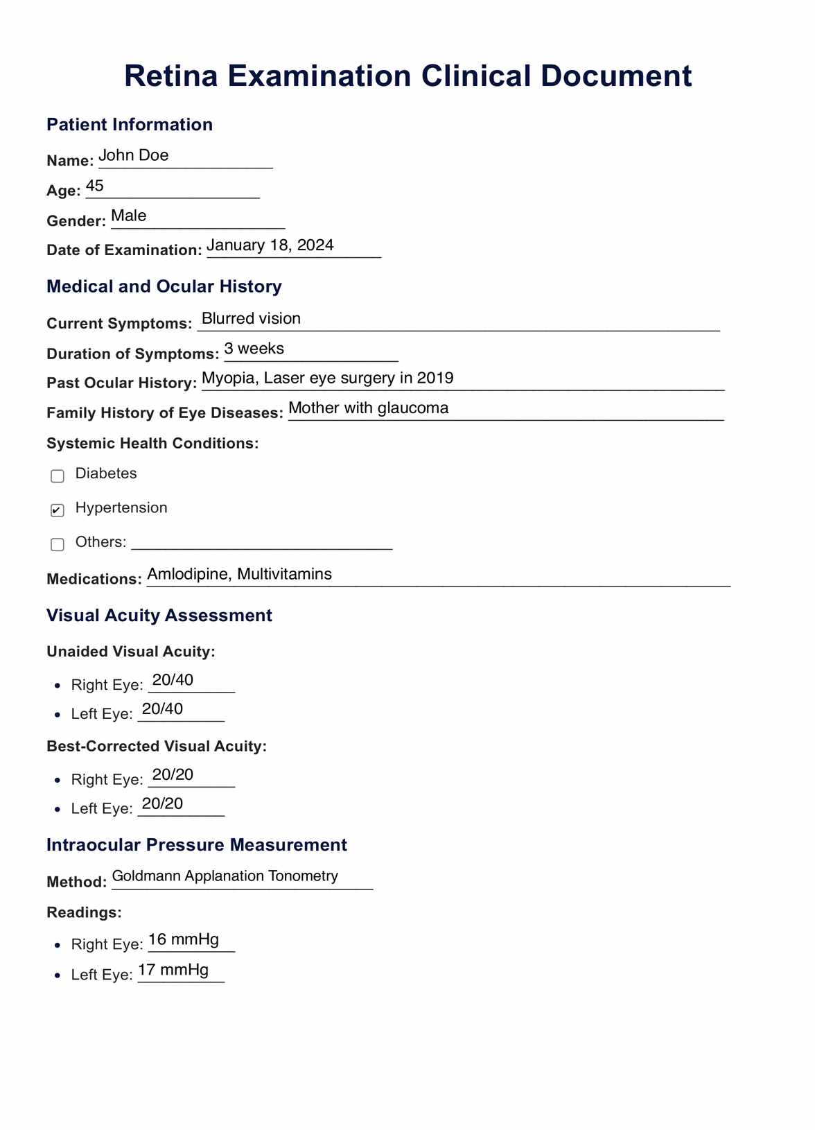
What is a Retina exam?
The retina, a crucial component of the eye, is a thin layer of tissue lining the back of the eye. Its primary function is to receive light that the lens has focused, convert this light into neural signals, and send these signals to the brain for visual recognition. The health of the retina is vital for clear vision.
A retina exam, often conducted by an ophthalmologist or an eye doctor, is a critical examination to assess the health of the retina and other structures within the eye. This type of eye exam is essential for early detection and treatment of various eye conditions and diseases that could lead to vision loss.
During a comprehensive eye exam, several tests are performed to evaluate the retina:
- Dilated eye exam: Eye drops are used to dilate the pupils, providing a clear view of the retina. This allows the eye doctor to examine the optic nerve, blood vessels, macula (the central part of the retina), and other structures inside the eye.
- Optical coherence tomography (OCT): This non-invasive imaging test provides cross-sectional pictures of the retina. It helps in detecting and treating retinal diseases like macular degeneration and diabetic retinopathy.
- Fluorescein angiography: A special dye (fluorescein dye) is injected into the bloodstream. The dye highlights the blood vessels in the back of the eye so they can be photographed. This test is particularly useful for diagnosing wet macular degeneration and other conditions involving abnormal blood vessels.
- Indirect ophthalmoscopy: This technique involves using a bright light and a special lens to examine the retina. It's especially useful for detecting retinal detachment, retinal tears, or other abnormalities.
- Visual field test: This test measures all areas of eyesight, including peripheral vision. It can help to detect abnormalities in the visual field caused by glaucoma and other eye diseases.
A retinal exam is crucial not only for those experiencing symptoms but also for individuals with risk factors such as diabetes, high blood pressure, or a family history of retinal diseases. Early detection through these exams can prevent conditions like diabetic retinopathy, which can lead to blindness if left untreated.
During the examination, the ophthalmologist may also look for signs of other eye conditions, such as glaucoma, by examining the optic disc and the pressure within the eye. The patient's retina is closely observed for any signs of damage, such as macular holes or retinal bleeding.
Regular eye exams, particularly for those with risk factors, are essential in maintaining good eye health and preventing vision loss. These exams allow doctors to monitor and treat any changes in the eye, ensuring the best possible outcome for the patient's vision.
Retina Exam Template
Retina Exam Example
When should you start getting your retina examined regularly?
The timing for when you should start getting your retina examined regularly depends on various factors such as your age, overall health, and risk factors for eye diseases. Here are some general guidelines:
- Adults with no significant risk factors:
- Age 20-39: An eye exam including a check of the retina every 5-10 years.
- Age 40-54: An eye exam every 2-4 years.
- Age 55-64: An eye exam every 1-3 years.
- Age 65 and older: An eye exam every 1-2 years.
- Individuals with risk factors:
- People with diabetes should have a dilated eye exam yearly since they are at higher risk for diabetic retinopathy.
- Those with a family history of eye diseases like glaucoma or macular degeneration should begin regular comprehensive eye exams in their 30s or earlier, based on their doctor's recommendation.
- Individuals with high blood pressure or other health conditions that can affect the eyes should follow their eye doctor's advice on how frequently to have their retinas checked.
- Children and adolescents:
- Children should have their first comprehensive eye exam at 6 months, then at age 3 and before first grade, as recommended by the American Optometric Association. While these exams focus more broadly on eye health and development, they can sometimes include a basic check of the retina.
- Symptoms or vision changes:
- Regardless of age or risk factors, if you experience any changes in vision such as blurriness, floaters, flashes of light, or loss of vision, you should schedule an eye exam immediately. These symptoms could indicate a retinal problem or other serious eye conditions.
- Specific recommendations:
- Your ophthalmologist or optometrist may have specific recommendations based on your individual eye health and family history. It's essential to follow their personalized advice.
It's important to remember that these are general guidelines, and the best course of action is to follow the advice of your eye care professional. Regular eye exams are crucial not only for vision health but also because they can detect other health issues.
What happens during a retinal eye exam?
During a retinal eye exam, various procedures are carried out to thoroughly examine the health of the retina and other parts of the eye. Here's an overview of what typically happens during such an examination:
- Patient history and symptoms discussion:
- The exam often begins with a discussion of your medical history, any symptoms you're experiencing, and any visual problems you've noticed. This might include questions about recent changes in your vision, family history of eye diseases, and any relevant health conditions like diabetes or high blood pressure.
- Visual acuity test:
- This test measures how well you can see at various distances. You'll be asked to read letters on a chart placed some distance away. This helps in assessing the clarity of your vision.
- Tonometry:
- This test measures the pressure inside your eye, which is important for detecting glaucoma. A device called a tonometer is used, sometimes after numbing the eyes with drops.
- Pupil dilation:
- To get a better view of the internal structures of your eye, especially the retina, the ophthalmologist will apply drops to dilate your pupils. Dilating the pupils allows more light into the eye and gives the doctor a clear view of the retina, optic nerve, and blood vessels.
- Ophthalmoscopy:
- This is a key part of the retinal exam. The eye doctor uses an ophthalmoscope, a device with a light and a magnifying lens, to examine the back of the eye, including the retina, optic disc, and blood vessels. This can be done through direct ophthalmoscopy (close up with a hand-held device) or indirect ophthalmoscopy (using a head-mounted device and a hand-held lens).
- Retinal imaging tests:
- Optical coherence tomography (OCT): This non-invasive imaging test provides detailed cross-sectional images of the retina, helping in the detection of conditions such as macular degeneration and diabetic retinopathy.
- Fluorescein angiography: In this test, a fluorescent dye is injected into a vein in the arm. The dye travels to the blood vessels in the eye, highlighting them to capture detailed images and detect problems like abnormal blood vessels.
- Visual field test:
- This test assesses your peripheral (side) vision. It can help in detecting eye diseases like glaucoma that can affect side vision.
- Slit lamp examination:
- The slit lamp is a microscope that allows the doctor to examine the eye under high magnification. While it's primarily used for the front parts of the eye, it can also provide views of the retina when used in conjunction with special lenses.
- Additional tests if needed:
- Depending on your specific situation, additional tests may be conducted to assess other aspects of your eye health.
After the examination, the eye doctor will discuss the findings with you. If any issues are detected, they will recommend a treatment plan or further testing. It's important to follow the doctor's advice and schedule follow-up appointments as necessary to monitor and manage any eye conditions.
Conditions that can be diagnosed through a retina exam
A retina exam is a critical tool for diagnosing a variety of eye conditions. Many of these conditions, if not identified and treated early, can lead to impaired vision or even blindness. Here are some of the key conditions that can be diagnosed through a retina exam:
- Diabetic retinopathy: This condition is a complication of diabetes, caused by damage to the blood vessels in the retina. It can lead to blood and fluid leakage, causing vision impairment.
- Macular degeneration: Age-related macular degeneration (AMD) affects the macula, the central part of the retina, leading to loss of central vision. It's one of the leading causes of vision loss in older adults.
- Retinal detachment: This is a medical emergency where the retina separates from its underlying layer. Symptoms can include flashes of light, floaters, and a shadow over your field of vision.
- Retinal vessel occlusion: This occurs when one of the vessels carrying blood to or from your retina becomes blocked, potentially leading to sudden vision loss.
- Glaucoma: While primarily an optic nerve disease, examining the retina can provide information about the progression of glaucoma, particularly in assessing any related damage to the retinal nerve fiber layer.
- Hypertensive retinopathy: Caused by high blood pressure, this condition can lead to changes in the retinal blood vessels, potentially resulting in blurred vision or loss of sight.
- Retinitis pigmentosa: This genetic disorder affects the retina and can lead to progressive vision loss.
- Macular hole: A small break in the macula, leading to blurred and distorted central vision.
- Epiretinal membrane: Also known as macular pucker, it involves the growth of a membrane that can distort vision.
- Central serous retinopathy: This condition involves fluid buildup under the retina, which can distort vision.
- Ocular tumors: Retinal exams can help in detecting tumors in the eye, like retinoblastoma in children or melanoma in adults.
- Retinopathy of prematurity: This is a condition in infants born prematurely, where abnormal blood vessels grow in the retina.
- Retinal hemorrhages: Bleeding in the retina, which can be a sign of various underlying conditions.
- Inherited retinal diseases: Conditions like Stargardt's disease or cone-rod dystrophy, which are genetic disorders affecting the retina.
A comprehensive retina exam not only helps in diagnosing these conditions but also in monitoring their progression and the effectiveness of treatment. Early detection through regular eye exams is key to preventing serious vision impairment or blindness associated with these conditions.
Treatment for patients with abnormal results
Treatment for patients with abnormal retinal exam results depends on the specific condition diagnosed. Here's an overview of treatments for various retinal conditions:
- Diabetic retinopathy:
- Laser surgery: Used to seal off leaking blood vessels and reduce swelling.
- Anti-VEGF injections: Medications like bevacizumab, ranibizumab, or aflibercept can help reduce swelling and growth of abnormal blood vessels.
- Vitrectomy: Surgical removal of the vitreous gel to treat severe bleeding or retinal detachment.
- Macular degeneration:
- Anti-VEGF injections: These medications help slow the progression of wet macular degeneration.
- Laser therapy: Certain types of laser treatment can help destroy actively growing abnormal blood vessels.
- Vitamins and supplements: Specific formulations can slow the progression of dry macular degeneration.
- Retinal detachment:
- Laser surgery or cryopexy: These procedures can repair a tear in the retina if caught early.
- Pneumatic retinopexy: A bubble of gas is injected into the eye to push the retina back into place.
- Vitrectomy or scleral buckle: Surgical procedures to reattach the retina.
- Retinal vessel occlusion:
- Laser treatment: To reduce swelling in the retina or to shrink abnormal new blood vessels.
- Medications: Injections to reduce macular edema.
- Glaucoma:
- Medicated eye drops: To reduce eye pressure.
- Laser treatment: To improve drainage of eye fluid.
- Surgery: Procedures like trabeculectomy or minimally invasive glaucoma surgeries (MIGS).
- Hypertensive retinopathy:
- Managing blood pressure: The primary treatment involves controlling high blood pressure.
- Macular hole and epiretinal membrane:
- Vitrectomy: Surgery to remove the vitreous and peel off the epiretinal membrane.
- Central serous tetinopathy:
- Often resolves on its own, but treatment options include laser therapy or photodynamic therapy in chronic cases.
- Ocular tumors:
- Treatment varies widely based on the type and stage of the tumor and can range from laser therapy and radiation to surgical removal.
- Retinopathy of prematurity:
- Laser therapy: To prevent abnormal blood vessel growth.
- Anti-VEGF injections: In certain cases.
- Inherited retinal diseases:
- Gene therapy: For specific genetic conditions like Leber's congenital amaurosis.
- Supportive care: Such as visual aids and rehabilitation.
In all cases, regular follow-up care is essential. The treatment approach should be tailored to the individual patient based on the specific diagnosis, severity of the condition, and overall health. Early detection and prompt treatment are key to preserving vision and preventing further deterioration.
Benefits of regular checkups
Regular eye checkups offer numerous benefits for maintaining both ocular and overall health. Here are some of the key advantages:
- Early detection of eye diseases:
- Many serious eye diseases, such as glaucoma, macular degeneration, and diabetic retinopathy, often have no early symptoms. Regular checkups can detect these conditions in their early stages, when they are most treatable.
- Prevention of vision loss:
- Early detection and treatment of eye diseases can prevent or delay vision loss. This is particularly important for conditions like glaucoma, where damage to vision is irreversible.
- Updating prescriptions:
- Regular checkups ensure that prescriptions for glasses or contact lenses are up to date. This is crucial for maintaining good vision and preventing vision-related problems like eye strain and headaches.
- Identifying systemic health issues:
- Eye doctors can detect signs of systemic health problems such as diabetes, high blood pressure, and high cholesterol during an eye exam. The eyes often reveal early signs of these conditions before other symptoms appear.
- Monitoring eye health over time:
- Regular checkups allow your eye doctor to track changes in your vision and eye health over time. This ongoing record can be invaluable in diagnosing and treating potential problems.
- Children’s vision development:
- Regular eye exams are crucial for children to ensure normal vision development. Eye problems in children can affect learning and development, so early detection and treatment are key.
- Promoting academic and professional performance:
- Good vision is essential for academic success and professional performance. Regular eye exams help ensure that vision issues do not hinder these areas of life.
- Enhancing quality of life:
- Good vision contributes significantly to overall quality of life. It enables you to enjoy daily activities, hobbies, and sports more fully.
- Eye comfort:
- Regular checkups can address issues like dry eyes, allergies, or eye strain, leading to greater comfort, especially for those who spend long hours in front of screens.
- Safety:
- Good vision is crucial for safety, especially in activities like driving. Regular eye exams help ensure that your vision meets the necessary standards for such activities.
In summary, regular eye checkups play a crucial role in maintaining your vision and eye health, detecting systemic diseases, and enhancing your overall quality of life. It's recommended to follow the eye exam schedule advised by your eye care professional based on your age, health status, and risk factors.
Commonly asked questions
A retinal exam helps eye doctors identify conditions like diabetic retinopathy and macular degeneration by examining the retina and optic nerve for abnormalities.
Optical coherence tomography (OCT) provides detailed images of the retina, crucial for diagnosing diseases like macular degeneration and assessing retinal thickness.
Abnormal blood vessels, often seen in diabetic retinopathy, are detected through dilated eye exams and tests like fluorescein angiography.
Related Templates

Popular Templates