The recovery time for a navicular stress fracture can vary, typically ranging from 6 to 8 weeks, depending on the severity of the fracture and the individual's overall health. It's crucial for recovery to follow a healthcare provider's advice closely, including rest and rehabilitation exercises. Some cases may require longer periods for full recovery, especially if surgical treatment is needed.
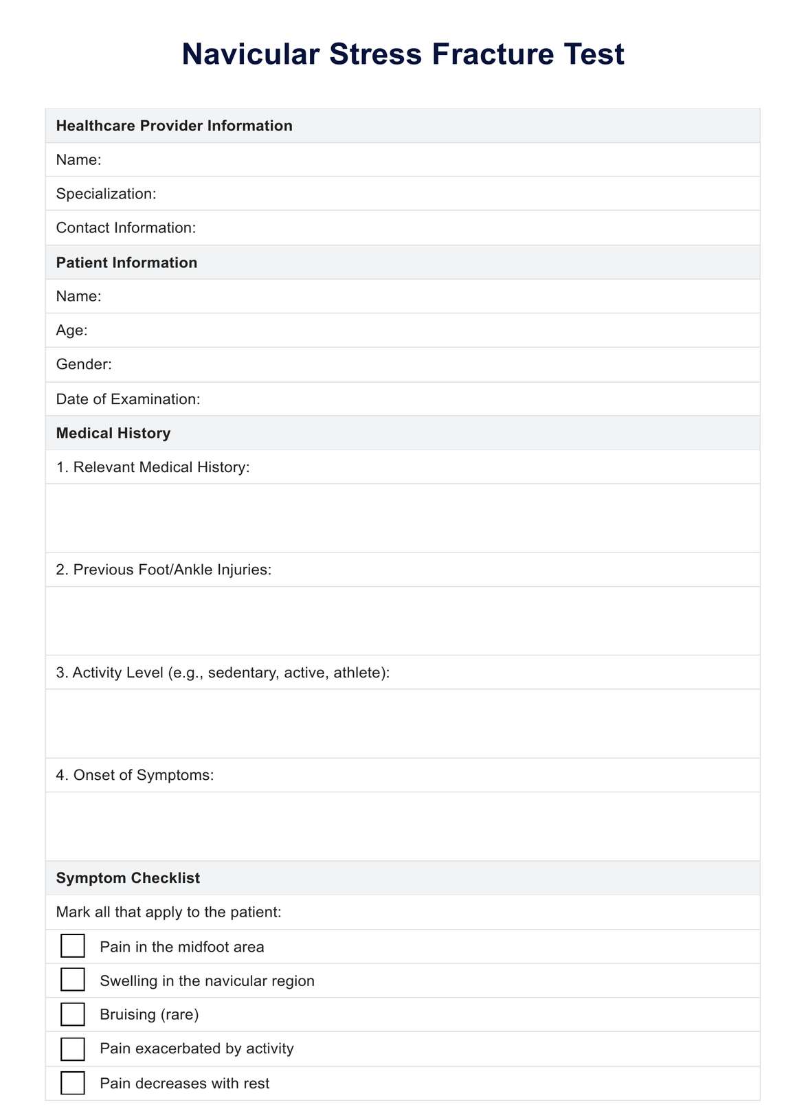
Navicular Stress Fracture Test
Explore diagnosis and treatment for navicular stress fractures with our free guide on tests, symptoms, and recovery strategies.

Use Template
Navicular Stress Fracture Test Template
Commonly asked questions
Walking on a navicular fracture is generally not recommended without professional advice. Doing so can aggravate the injury and prolong the healing process. Healthcare providers often suggest using crutches or a walking boot to offload weight from the foot until it's safe to bear weight again. Always consult with a healthcare provider for guidance on weight-bearing activities during recovery.
Whether a navicular fracture needs a cast depends on the fracture's severity and location. Many navicular fractures, especially stress fractures, are treated with non-surgical methods, including immobilization with a cast or a protective boot to limit movement and aid in healing. Surgical treatment may be required in more severe cases, followed by casting. A healthcare provider will determine the best treatment plan based on individual assessments.


EHR and practice management software
Get started for free
*No credit card required
Free
$0/usd
Unlimited clients
Telehealth
1GB of storage
Client portal text
Automated billing and online payments