Leg Length Discrepancy Test
Discover the Leg Length Discrepancy Test: Measure and address differences in leg length for better patient outcomes. Download our free PDF guide now.
What is Leg Length Discrepancy?
Leg length discrepancy (LLD), also known as anisomelia or leg length asymmetry, refers to having unequal leg lengths. This disparity can stem from a congenital leg length discrepancy, where the condition is present at birth. LLD can be structural or functional, arising from body asymmetries like pelvic tilt.
True leg length discrepancy vs. functional leg length discrepancy
When discussing leg and limb length discrepancies above, it's important to distinguish between true leg length discrepancy and functional leg length discrepancy:
- True leg length discrepancy: This type, also known as structural leg length discrepancy, involves an actual, measurable difference in the bone lengths of the legs, sometimes referred to as limb length difference or limb length inequality. It can be caused by congenital growth abnormalities or idiopathic leg length discrepancy, which occurs without a known cause.
- Functional leg length discrepancy: Functional LLD, not involving actual differences in limb length, arises from conditions like pelvic tilt or muscular imbalances, leading to a perceived limb shortening. This could be due to issues like muscle imbalances, abnormal posture, or other conditions that affect how the legs are used and positioned, such as pelvic tilt.
Potential problems leg length discrepancy may lead to
Whether true or functional, LLD can lead to various issues affecting overall health and mobility. Some of the potential problems include:
- Gait abnormalities: Individuals with LLD often develop abnormal walking patterns, exacerbating issues like short leg syndrome. This might manifest as limping, toe walking on the shorter side, or bending the knee on the longer side.
- Postural issues: Having a shorter leg can lead to many problems, including chronic pain. This misalignment can extend upwards, affecting the spine and eventually leading to scoliosis or kyphosis.
- Increased risk of injury: The compensatory mechanisms and altered biomechanics associated with leg length differences can increase the risk of injuries, particularly in the lower back, hips, and knees. A study by Rauh in 2018 reported that people with LLD above 1.5 cm are at higher risk of lower leg injuries.
- Chronic pain: Chronic pain in the back, hips, knees, or ankles can develop due to the continuous stress and strain of compensating for a leg length discrepancy.
- Reduced physical performance: A minor leg length difference can significantly impact athletes' performance. The imbalance can affect running efficiency, agility, and balance, potentially hindering athletic performance and increasing the risk of sports-related injuries.
How to diagnose Leg Length Discrepancy
Diagnosing LLD involves a combination of physical examination and precise leg length measurement techniques.
Healthcare providers begin with a physical exam as a screening by observing the patient's gait and looking for signs of limb length discrepancy or imbalance. This assessment has multiple options; the examiner may choose between direct measurement using a tape measure (known as the direct method), palpation of bony landmarks (known as the indirect method), or the Palpation Meter (PALM) method.
Next, the examiner may use imaging tests to provide detailed images of the bones and soft tissues as well as accurate measurements. In addition, attention is paid to any functional leg length discrepancies, which may not involve actual differences in bone length but result from alignment issues in the pelvis or lower extremities.
Leg Length Discrepancy Test Template
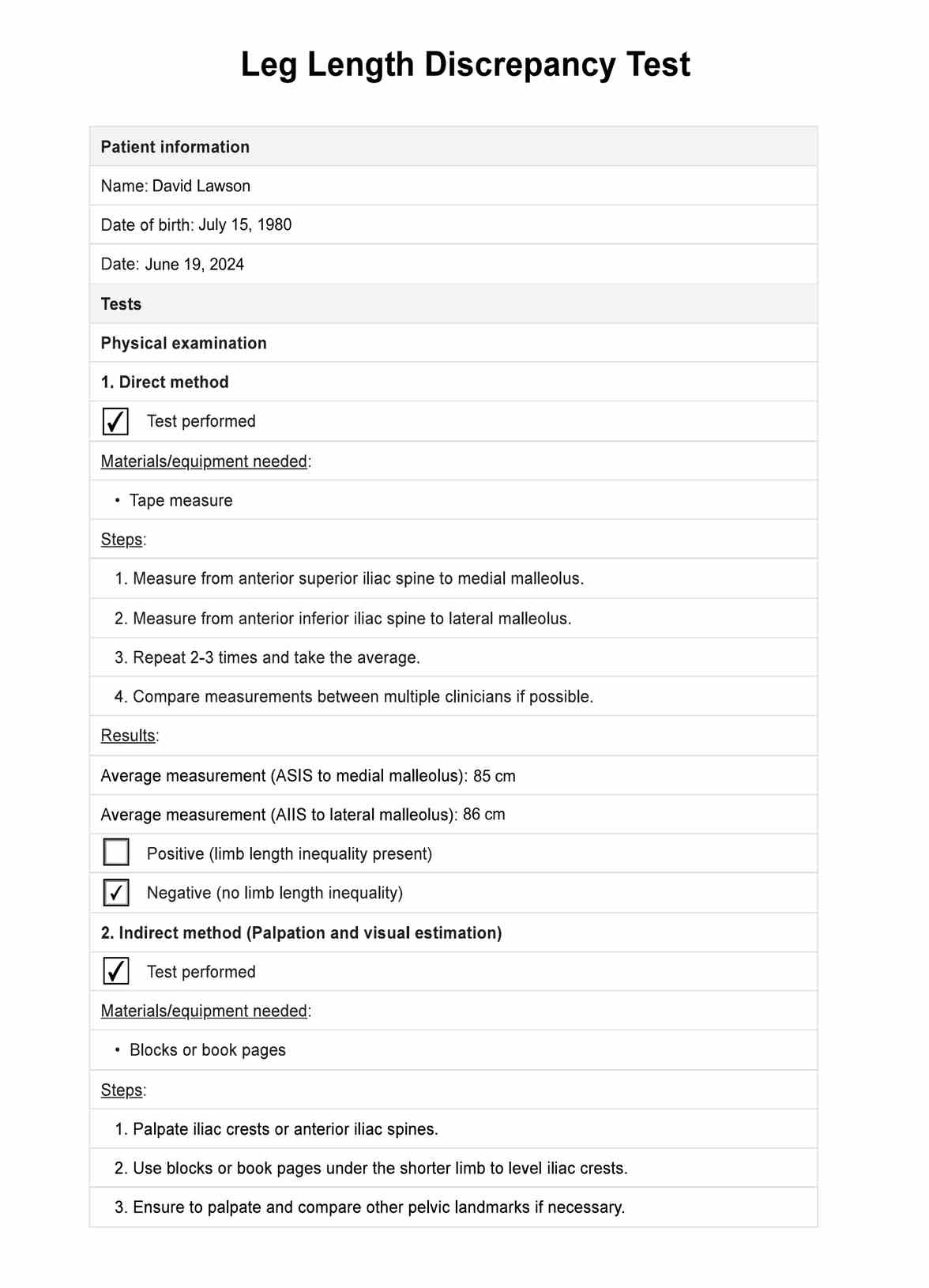
Leg Length Discrepancy Test Example
Different Leg Length Discrepancy Tests
To accurately diagnose and quantify the extent of a leg length discrepancy, healthcare professionals can employ several tests. Here are three widely used methods to assess leg length discrepancies:
Physical examination
In a physical examination, healthcare professionals measure leg length through various methods . This method involves both direct and indirect techniques, as well as the use of specialized tools like the PALM (Palpation Meter).
Direct method
The direct method involves measuring limb length with a tape measure between two defined points while the patient is standing. Common points include the anterior superior iliac spine and the medial malleolus or the anterior inferior iliac spine and the lateral malleolus. To enhance accuracy, it is recommended to use the average of at least two or three measurements. Compare measurements between two or more clinicians when possible.
However, tape measure methods have potential sources of error, such as iliac asymmetries, unilateral deviations (e.g., genu varum), asymmetrical umbilical position, and joint contractures, which can mask or exaggerate limb length discrepancies.
Indirect method (palpation and visual estimation)
The indirect method involves palpating bony landmarks, typically the iliac crests or anterior iliac spines, while the patient is standing. Clinicians detect if these landmarks are at the same horizontal level. This technique often uses blocks or book pages of known thickness under the shorter limb to adjust until the iliac crests are level. It's crucial to consider asymmetric pelvic rotations in other planes, as they may affect the perceived limb length inequality. To increase precision, clinicians should palpate and compare as many pelvic landmarks as possible, including the greater trochanter major.
Palpation meter (PALM)
The PALM is a reliable and valid instrument for measuring pelvic height differences. It is a cost-effective alternative to radiographic measurements. The procedure involves these steps:
- Place two tape strips on the ground, 15 cm apart, where patients align the medial borders of their feet.
- Ask patients to walk for 10 steps and stand fully erect (no bending of the knee, hip, or spine).
- Place the PALM on the most superior aspect of the iliac crest and measure the distance between caliper heads to the nearest millimeter and the angle of inclination to the nearest half-degree. The inclinometer ball will move towards the side of the shorter limb.
Block test
The Block Test is a straightforward clinical method for determining the extent of leg length discrepancy. Here's how to do it:
- Have the patient stand with their feet 10 cm apart, knees extended, and weight equally distributed on both feet.
- Place your hands on bilateral anatomical landmarks such as the spina iliaca posterior superior, spina iliaca anterior superior, or crista iliaca.
- Visually assess if there is a length inequality by checking the level of the hips and shoulders.
- If inequality is detected, place a wooden board of 0.5 cm thickness under the foot of the shorter side.
- Continue to add thicker planks under the shorter limb until the hips and shoulders appear level.
- The thickness of the final plank stack used to level the patient is equal to the leg length discrepancy.
This test has factors such as pelvic asymmetry, incorrect foot positioning, obesity, joint contractures, scoliosis, and measurement inaccuracies.
Imaging and radiographic measurement
For a more detailed and accurate analysis, physicians may use various imaging methods, which are considered the gold standard (Sabharwal & Kumar, 2008). These methods include plain radiography techniques such as orthoroentgenogram, scanogram, and teleoroentgenogram, which provide full-length standing AP radiographs of the lower extremity.
Computed radiography and micro-dose digital radiography are also employed, with the latter significantly reducing radiation exposure compared to traditional radiographic techniques. Ultrasound imaging uses a transducer to locate bony landmarks at the hip, knee, and ankle joints. CT scanograms offer anteroposterior (AP) scout views of the femurs and tibias. MRI scans provide detailed images using a T1 weighted spin echo sequence, selecting the best coronal images for standardized assessment of femoral length based on classic bony landmarks such as the femoral head and medial femoral condyle.
After a discrepancy is measured, you may take different courses of action depending on severity. For example, a discrepancy of 0 to 2 cm may not require treatment, while a discrepancy of 2 to 6 cm may be treated with shoe lifts, epiphysiodesis, or shortening. Larger discrepancies may require more complex procedures such as lengthening. These options will be discussed in a later section.
Treatment of Leg Length Discrepancy
Treatment options for leg length inequality depend on the severity of the limb length difference and the patient's symptoms. Here's a breakdown of the most common approaches:
- Shoe lifts: For mild to moderate discrepancies, shoe lifts or heel inserts can balance the legs to an equal length. This can help improve alignment and relieve pain caused by the discrepancy.
- Physical therapy: Specifically tailored exercises can enhance muscle strength and improve posture. Physical therapy can strengthen the muscles around the lower back, hips, and legs, helping improve mobility and reduce pain. It may also address compensatory behaviors that might develop from the discrepancy.
- Surgical options: In cases of significant discrepancy, especially those over 2 cm or where non-surgical methods have not provided relief, surgery might be considered. Surgical methods can vary:
- Epiphysiodesis: This is a procedure used primarily in children, which involves halting the growth of the longer leg to allow the shorter leg to catch up.
- Leg lengthening: Also known as limb lengthening, this involves surgically breaking the bone and using a device to gradually stretch the bones as they heal, allowing the shorter leg to lengthen.
- Bone resection: Surgery may involve removing a section of the bone in the longer leg for certain types of discrepancies to even out the length.
Each treatment strategy is selected based on the specific needs of the individual, focusing on minimizing the physical and functional impacts of LLD.
References
Rauh, M. J. (2018). Leg-length inequality and running-related injury among high school runners. International Journal of Sports Physical Therapy, 13(4), 643–651. https://www.ncbi.nlm.nih.gov/pmc/articles/PMC6088132/
Sabharwal, S., & Kumar, A. (2008). Methods for assessing leg length discrepancy. Clinical Orthopaedics and Related Research, 466(12), 2910–2922. https://doi.org/10.1007/s11999-008-0524-9
Commonly asked questions
It is diagnosed through physical examinations, measurements of leg lengths from hip to ankle, and imaging tests like X-rays or MRIs to confirm the extent of the discrepancy.
Radiographic measurements are generally considered the most precise method to assess leg length discrepancies accurately.
The best imaging modality for accurately assessing leg length discrepancy (LLD) is full-length standing anteroposterior radiography, also known as a scanogram or orthoroentgenogram. Compared to other imaging modalities like CT or MRI, scanograms provide the necessary full-length visualization of the lower limbs while minimizing radiation exposure and cost.