Stress Fracture Diagnosis Worksheet

Our Stress Fracture Diagnosis Worksheet aids healthcare professionals in thorough assessment and documentation for accurate diagnosis and treatment planning of stress fractures.

By Bernard Ramirez on Jun 30, 2024.

tick

Fact Checked by Ericka Pingol.

Use Template
Stress Fracture Diagnosis Worksheet PDF Example
toolsShare uiAI icon

How do you know if someone has a stress fracture?

Detecting a stress fracture involves a thorough assessment. Early signs of stress fractures include pain and tenderness in weight-bearing bones, often exacerbated by physical activity. Patients may initially experience pain during activity, progressing to constant discomfort, even at rest.

To diagnose stress fractures, healthcare professionals rely on a combination of clinical evaluation and imaging. Magnetic resonance imaging (MRI) is the preferred method due to its high sensitivity and ability to detect early tibial stress injuries and stress reactions before they fully develop into fractures. MRI is particularly useful in diagnosing medial tibial stress syndrome, a condition that precedes stress fractures in the tibia.

Most stress fractures occur in athletes and military personnel who engage in repetitive, high-impact activities. Relieving pain through rest and modifying physical activity levels is crucial for recovery. Early diagnosis and intervention are essential to prevent injury worsening and facilitate a quicker return to normal activities.

Printable Stress Fracture Diagnosis Worksheet PDF

Download this Stress Fracture Diagnosis Worksheet to help in accurate diagnosis and effective management of stress fractures, ensuring optimal patient care and outcomes.

Stress Fracture Diagnosis Worksheet example (sample)

Our Stress Fracture Diagnosis Worksheet PDF is a comprehensive tool for healthcare professionals. It guides thorough patient assessments, including history, physical exam, and imaging tests like X-rays and bone scans. This template aids in accurate diagnosis and effective management of stress fractures, ensuring optimal patient care and outcomes.

Download our free Stress Fracture Diagnosis Worksheet example here

Stress Fracture Diagnosis Worksheet example (sample)

How do people develop stress fractures?

Stress fractures develop due to repetitive stress and overuse of bones, particularly in athletes and military personnel. Female athletes are at a higher risk due to hormonal imbalances and lower bone density. These fractures result from an imbalance between bone breakdown and repair, often exacerbated by sudden increases in training intensity, improper footwear, and biomechanical abnormalities.

Several modifiable risk factors contribute to the development of stress fractures, including an abrupt increase in physical activity, improper training techniques, and inadequate nutrition. A well-designed exercise program can mitigate these risks by promoting gradual increases in intensity and ensuring proper technique.

Clinically, a stress fracture feels like localized pain that worsens with activity and improves with rest. During a physical exam, healthcare providers look for tenderness over the injured bone and may use musculoskeletal imaging to confirm the diagnosis. Bone scintigraphy findings can also be useful in detecting stress fractures, especially when other imaging modalities are inconclusive.

How do nurses diagnose stress fractures?

Nurses play a crucial role in the initial diagnosis and management of stress fractures. To diagnose stress fractures, nurses conduct thorough patient assessments and consider risk factors such as eating disorders, recent increases in physical activity, and biomechanical abnormalities that contribute to bone stress injury.

Assessment

Nurses begin by collecting a detailed patient history, focusing on symptoms, physical activity levels, and underlying conditions. They look for signs of a tibial stress fracture or other bone stress injuries, such as localized pain that worsens with activity and improves with rest.

Physical examination

A physical exam helps identify tenderness and swelling at the injury site. Nurses assess for signs of bone stress reactions, indicating the potential for developing stress fractures.

Imaging tests

Nurses often coordinate with physicians to order appropriate imaging tests to confirm a stress fracture diagnosis. Bone scans and magnetic resonance imaging (MRI) are particularly useful. Bone scans detect increased metabolic activity indicative of a stress reaction, while MRI provides detailed images to identify bone stress injuries early.

Treatment and prevention

Once a stress fracture is diagnosed, nurses educate patients on stress fracture treatment, including rest, activity modification, and sometimes protective footwear or braces to ensure the injury heals properly. They emphasize the importance of nutrition and gradual increases in physical activity to prevent stress fractures.

Can you diagnose a stress fracture without magnetic resonance imaging (MRI)?

Diagnosing stress fractures without MRI requires a comprehensive approach. A patient's history and physical examination are crucial in identifying tenderness, swelling, and signs of bone stress reactions. X-rays often identify more developed fractures, while bone scans detect increased metabolic activity.

Computed tomography (CT) can provide detailed images of the bone but is less sensitive to early stress reactions. Ultrasound can detect changes in the bone or surrounding tissues associated with stress fractures. Clinical management focuses on stress fracture treatment, including rest, activity modification, and addressing risk factors to ensure proper healing and prevent recurring fractures.

How is a stress fracture treated?

Treating a stress fracture involves a comprehensive approach to ensure proper healing and prevent recurrence. Here is a detailed look at how stress fractures are treated:

Rest and activity modification

The cornerstone of stress fracture treatment is rest. Patients must significantly reduce or completely stop activities that cause pain. This allows the injured bone to heal properly and prevents further damage.

Pain management

To relieve pain, nonsteroidal anti-inflammatory drugs (NSAIDs) may be recommended. Ice application can also reduce swelling and alleviate discomfort.

Nutritional support

Ensuring adequate nutrition is vital, particularly for patients with eating disorders or nutritional deficiencies. A balanced diet rich in calcium and vitamin D supports bone healing and overall bone health.

Immobilization

In some cases, immobilization with a brace or walking boot may be necessary. This helps stabilize the injured bone and reduce stress during the healing process.

Physical therapy

Once initial pain subsides, physical therapy can help restore strength and flexibility. A tailored exercise program gradually reintroduces weight-bearing activities, promoting a return to normal function.

Gradual return to activity

A key aspect of treatment is a carefully monitored return to physical activity. This involves gradually increasing the intensity and duration of exercise to prevent stress reactions and future stress fractures.

Advanced interventions

Surgical intervention might be necessary in severe cases where conservative treatment is insufficient. Internal fixation with pins or screws can stabilize the fracture and promote healing.

Prevention strategies

Educating patients on how to prevent stress fractures is vital. This includes maintaining a balanced diet, ensuring gradual increases in physical activity, and using appropriate training techniques.

Why use Carepatron as your physical activity tracking software?

Carepatron is a specialized EMR for physical therapy and rehabilitation, offering comprehensive features for healthcare professionals. It allows for customization and integration of treatment plans and exercise programs to each patient's specific needs, integrating patient goals, progress tracking, and outcomes assessment into the clinical workflow.

Carepatron promotes active patient engagement through its user-friendly interface and patient portal capabilities, fostering a collaborative approach to care.

The software excels in clinical documentation, capturing detailed notes, assessments, and treatment outcomes, ensuring thorough documentation of physical activity tracking. Its analytics tools provide insights into patient progress, adherence to treatment plans, and outcomes, aiding in evidence-based decision-making and continuous quality improvement.

As a HIPAA-compliant platform, Carepatron prioritizes patient data security and confidentiality, safeguarding sensitive health information. It seamlessly integrates with existing healthcare systems and devices, ensuring continuity of care across different settings.

Practice Management Software
What is the Stress Fracture Diagnosis Worksheet used for?

Commonly asked questions

What is the Stress Fracture Diagnosis Worksheet used for?

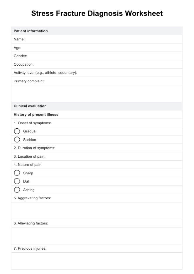
The worksheet is used to assess patients suspected of stress fractures systematically. It includes sections for patient history, physical examination findings, and imaging results to aid in diagnosis.

Why is it important to use this worksheet?

Using the worksheet ensures a thorough evaluation, helping healthcare providers identify stress fractures early. It standardizes the diagnostic process, ensuring comprehensive care and accurate treatment planning.

What information does the worksheet include?

The worksheet includes patient demographics, details of symptoms, risk factors, physical exam findings such as tenderness and swelling, and results from imaging tests like X-rays or bone scans.

Join 10,000+ teams using Carepatron to be more productive

One app for all your healthcare work