Prenatal Blood Test
Explore our comprehensive guide on prenatal blood tests and ultrasounds, offering vital insights for healthcare professionals. Download our free PDF now!
What is prenatal blood testing?
Prenatal blood testing is vital in the pregnancy journey, offering a window into the health and well-being of both mother and unborn child. As healthcare professionals, you play a crucial role in this journey, guiding expectant mothers through the maze of prenatal care with expertise and compassion.
This type of testing, typically conducted in the first trimester, involves analyzing a pregnant woman's blood to assess various health indicators. These indicators can include blood type, Rh factor, and levels of certain substances, which may provide early insight into potential genetic disorders or developmental issues in the fetus. It's not just about detecting problems; it's also about peace of mind and preparation. Knowing what's normal and what's not can help you, as a healthcare provider, offer better, more personalized care to your patients.
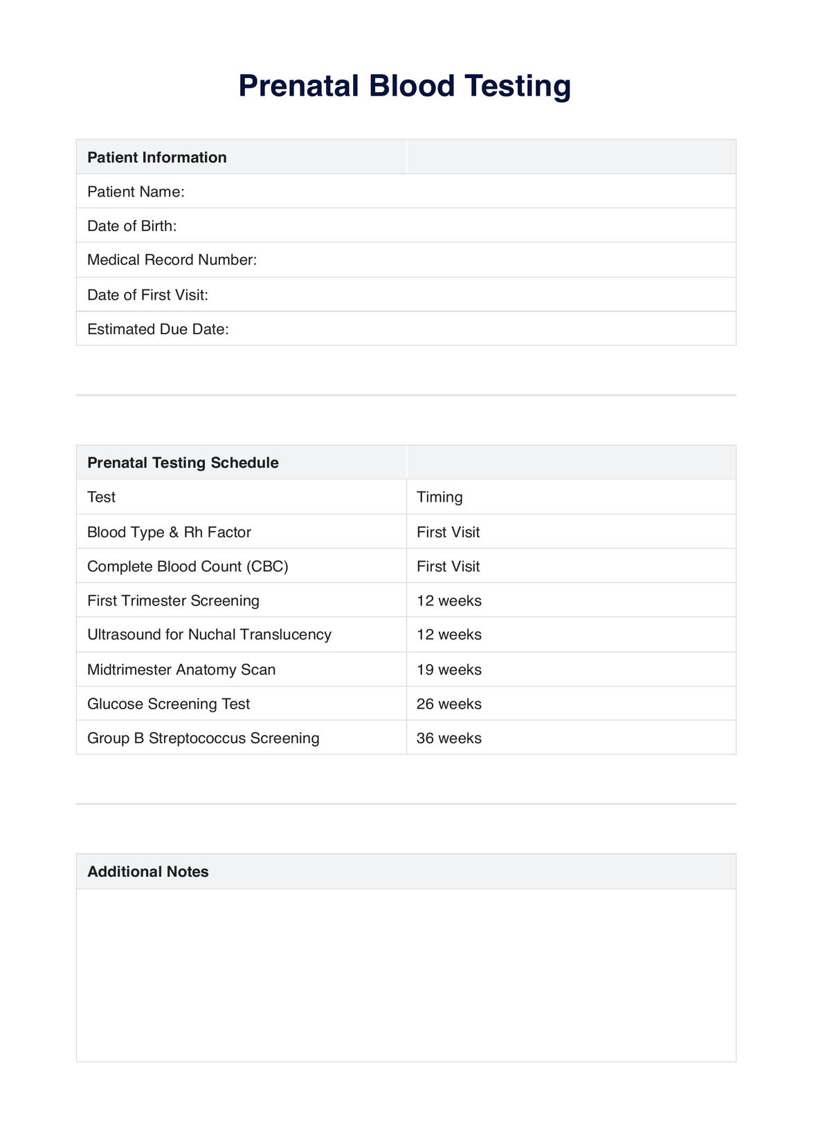
Our comprehensive guide delves into the specifics of prenatal blood testing, providing examples and detailed explanations to enhance your understanding. This resource is designed to be a valuable tool in your practice management software, aiding in informed decision-making and patient education.
Our guide is more than just information; it's a support system for healthcare professionals like you, who are committed to offering the best prenatal care. With this knowledge, you can confidently navigate the complexities of prenatal testing, ensuring the health and safety of both mother and child.
Download our free PDF to explore this crucial aspect of prenatal care in more detail. It's not just a guide; it's a resource created to support you as a caring and authoritative figure in the healthcare industry.
Prenatal Blood Test Template
Prenatal Blood Test Example
Types of prenatal testing
Prenatal testing is a multifaceted approach, encompassing various tests conducted at different stages of pregnancy. Each diagnostic test serves a unique purpose, providing crucial information about the fetus's and the mother's health. Let's delve into the specifics of these tests.
First-trimester prenatal screening tests
The first trimester, a critical period in pregnancy, involves several key screening tests. These tests are designed to evaluate the various risk factors of the fetus having specific chromosomal abnormalities, such as Down syndrome (trisomy 21) or Edwards syndrome (trisomy 18).
- Blood tests: These measure levels of specific substances in the mother's blood, which can indicate the likelihood of chromosomal abnormalities.
- Ultrasound for nuchal translucency: This ultrasound measures the thickness of the fluid at the back of the baby's neck. An increased thickness can indicate a higher risk of specific congenital disabilities.
- Combined first-trimester screening: This combines the blood tests and the nuchal translucency ultrasound to provide a more comprehensive risk assessment.
These screenings don't diagnose conditions; they assess risk levels, guiding further diagnostic or genetic testing, if necessary.
Mid-trimester (also called the 18- to 20-week scan)
The mid-trimester scan, often called the anatomy scan, is a detailed ultrasound performed between the 18th and 20th weeks of pregnancy. This scan is pivotal in assessing the physical development of the fetus. Key aspects of this scan include:
- Assessment of fetal anatomy: Check the development of the fetus's organs and detect any abnormalities.
- Measurement of fetal size: Ensuring the fetus is growing at a healthy rate.
- Placental position and amniotic fluid volume: Evaluating the placenta's position and the amount of amniotic fluid surrounding the baby are crucial for fetal health.
- Gender determination: If desired, the baby's sex can often be identified during this scan.
This scan is a comprehensive check-up, offering reassurance and, in some cases, critical information for managing potential complications.
When are ultrasounds performed during pregnancy?
Ultrasounds are a routine part of prenatal care and are performed at various stages of pregnancy to monitor the development of the fetus and the health of the mother. The timing of these ultrasounds can vary, but generally, they are scheduled as follows:
- First trimester (6-12 weeks): Early ultrasound to confirm the pregnancy, estimate the due date, and check for multiples (twins, triplets, etc.).
- Nuchal translucency scan (11-13 weeks): Part of the first-trimester screening for chromosomal abnormalities.
- Anatomy scan (18-20 weeks): A detailed scan to examine the fetus's anatomy, including the brain, heart, kidneys, and limbs.
- Third-trimester ultrasounds (28-40 weeks): These may be performed to monitor the growth of the fetus, the amount of amniotic fluid, and the position of the placenta.
Additional ultrasounds may be recommended based on the specific needs of the pregnancy, such as in cases of increased risk of high-risk pregnancies, if there are concerns about fetal growth, or if complications arise.
What are the risks and benefits of ultrasound imaging?
While considered safe, ultrasound imaging has risks and benefits that healthcare providers and expectant mothers should understand.
Benefits
- Safe for mother and baby: Ultrasound uses sound waves, not radiation, making it safe for both the mother and the fetus.
- Diagnostic tool: It helps diagnose and monitor various aspects of fetal development and maternal health.
- Detection of abnormalities: Early detection of potential issues can lead to timely interventions.
- Emotional bonding: Seeing the fetus can help parents start bonding with their unborn child.
Risks
- False positives/negatives: Ultrasound can sometimes give false positive or negative readings, leading to unnecessary stress or reassurance.
- Overdiagnosis: It may detect minor issues that are not clinically significant, leading to unnecessary interventions.
- Physical discomfort: While usually painless, some women may find the procedure physically uncomfortable.
In conclusion, while ultrasounds are an invaluable tool in prenatal care, healthcare providers need to use them judiciously and discuss their limitations with patients.
Interpreting the results
Interpreting the results of prenatal tests is a critical aspect of prenatal care. For healthcare professionals, it's essential to understand what the results indicate and how to communicate them effectively to expectant mothers.
- Normal results: These typically indicate that the mother and fetus are healthy and the pregnancy is progressing normally. It's essential to reassure the patient and continue with routine prenatal care.
- Borderline results: Sometimes, results may fall into a borderline category. In such cases, it may be necessary to repeat the test or perform additional tests for a more definitive understanding.
- Abnormal results: Abnormal findings can be a source of anxiety for patients. It's crucial to provide clear, empathetic communication, explaining what the results mean and that an abnormal result does not always indicate a problem.
Healthcare providers should ensure patients understand their results and feel supported, regardless of the outcome.
Next steps for patients with abnormal findings
When prenatal tests yield abnormal results, it's essential to have a clear plan of action:
- Detailed explanation: Begin by providing a detailed explanation of what the abnormal results mean. It's important to be factual yet sensitive in delivering this information.
- Further testing: Often, abnormal results require further testing for confirmation. This may include additional blood tests, specialized ultrasounds, or diagnostic tests like amniocentesis or chorionic villus sampling (CVS).
- Referral to specialists: Depending on the findings, referral to a genetic counselor or a maternal-fetal medicine specialist may be necessary.
- Support and counseling: Offer emotional support and counseling. Receiving abnormal results can be stressful, and patients may need help processing the information and understanding the implications.
- Developing a care plan: Work with the patient to create one. This may involve more frequent monitoring, changes in lifestyle, or even medical intervention, depending on the nature of the findings.
Healthcare providers must guide patients through this process with empathy and expertise, ensuring they receive the appropriate healthcare provider and support.
Research and evidence
The utilization of prenatal blood testing and ultrasound imaging in prenatal care is supported by extensive research and evidence, underscoring their critical role in ensuring maternal and fetal health.
A study by Lunardhi et al. (2023) Predictive Ability of Ultrasound on Neonatal Diagnoses and Consequences on Prenatal Care Utilization highlights the accuracy of fetal ultrasounds in predicting neonatal diagnoses. The research at Harbor-UCLA Medical Center analyzed cases with abnormal fetal ultrasound findings and their correlation with neonatal outcomes. The study found significant associations between ultrasound findings and neonatal conditions like congenital heart disease and growth restriction, though it also noted high false-positive rates for specific diagnoses. This emphasizes the importance of understanding the limitations of ultrasound in prenatal care.
In another pivotal study by Salomon et al. (2017), ISUOG updated the consensus statement on the impact of cfDNA aneuploidy testing on screening policies and prenatal ultrasound practice and discussed integrating cell-free DNA (cfDNA) testing with traditional ultrasound practices. The research underscores the need to balance cfDNA testing with ultrasound to provide comprehensive prenatal care, highlighting the evolving landscape of prenatal screening.
Kelly-Hedrick et al. (2022), in their work Perceived Value of Prenatal Ultrasound Screening: A Survey of Pregnant Women, explored pregnant women's perceptions regarding prenatal ultrasounds. The study revealed that women value ultrasound screenings for preparedness and knowledge beyond their clinical decision-making utility. This insight is crucial for healthcare providers in understanding patient perspectives and enhancing prenatal care.
These studies demonstrate the critical role and evolving nature of prenatal testing in healthcare, providing a foundation for current practices and future advancements in prenatal care.
References
Kelly-Hedrick, M., Geller, G., Jelin, A., & Gross, M. (2022). Perceived Value of Prenatal Ultrasound Screening: A Survey of Pregnant Women. Maternal and Child Health Journal. https://dx.doi.org/10.1007/s10995-022-03515-1
Lunardhi, A., Goldrich, A., Stohl, H., Ko, K. J., Dong, F., & Galyon, K. D. (2023). Predictive Ability of Ultrasound on Neonatal Diagnoses and Consequences on Prenatal Care Utilization. Ultrasound Quarterly. https://dx.doi.org/10.1097/RUQ.0000000000000665
Salomon, L., Alfirevic, Z., Audibert, F., Kagan, K., Paladini, D., Yeo, G. S. H., & Raine-Fenning, N. (2017). ISUOG updated consensus statement on the impact of cfDNA aneuploidy testing on screening policies and prenatal ultrasound practice. Ultrasound in Obstetrics & Gynecology. https://dx.doi.org/10.1002/uog.17483
Toscano, M., Marini, T., Drennan, K., Baran, T., Kan, J., Garra, B., Dozier, A., Ortega, R., Quinn, R., Zhao, Y. T., Egoavil, M., Tamayo, L., Carlotto, C., & Castañeda, B. (2021). Testing telediagnostic obstetric ultrasound in Peru: a new horizon in expanding access to prenatal ultrasound. BMC Pregnancy and Childbirth. https://dx.doi.org/10.1186/s12884-021-03720-w
Commonly asked questions
Prenatal blood testing is a series of blood tests conducted during pregnancy to assess the health of both the mother and the fetus. These tests can detect genetic conditions and potential issues, ensuring timely interventions.
The first ultrasound in pregnancy is typically performed between 6 to 12 weeks. This early scan is crucial for confirming the pregnancy, estimating the due date, and checking for multiple pregnancies.
Prenatal ultrasounds are generally considered safe and non-invasive. They use sound waves to create images of the fetus and do not involve radiation. However, it's essential to use them judiciously and under the guidance of a healthcare professional.