Kidney Stone Analysis
Learn more about Kidney Stone Analysis. Download our free PDF template to record your patient's information and streamline clinical documentation.


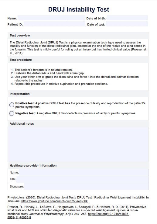
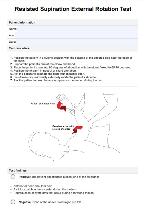
What is a Kidney Stone Analysis?
A Kidney Stone Analysis is a medical diagnostic procedure involving a detailed examination of a kidney stone to determine its composition and structure. Kidney stones are small, hard mineral deposits that can form in the kidneys and can be extremely painful when they pass through the urinary tract.
The analysis of these stones is crucial for identifying the composition of a kidney stone and is essential for determining the most appropriate treatment. Different types of kidney stones require different approaches, such as dietary modifications, medication, or surgical intervention.
Understanding the composition of a kidney stone can also help healthcare professionals advise patients on how to prevent future stone formation. Dietary and lifestyle recommendations can be tailored to reduce the risk of recurrence.
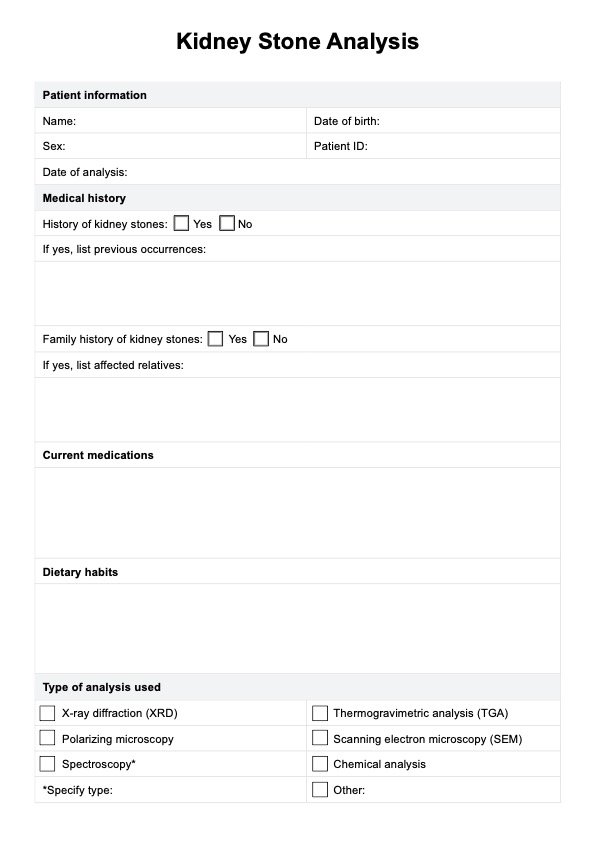
Kidney Stone Analysis Template
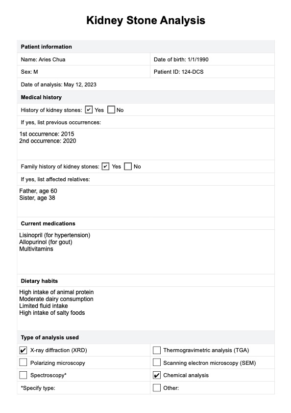
Kidney Stone Analysis Example
How does our Kidney Stone Analysis template work?
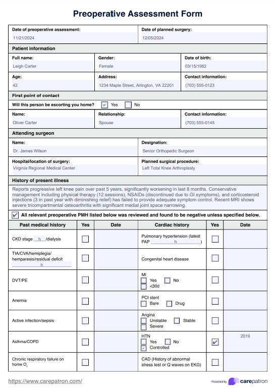
Our Kidney Stone Analysis template is designed to simplify the documentation process for healthcare professionals. It includes sections for recording essential patient information, medical history, and the type of analysis performed. Here's how to use the template:
Step 1: Download the resource
Click "Use Template" to utilize the Kidney Stone Analysis form via the Carepatron app. Choose "Download" for a PDF copy.
Step 2: Fill in patient information
Start by entering the patient's name, date of birth, sex, patient ID, and the date of the analysis. This ensures that all critical identifying information is captured before beginning the assessment.
Step 3: Document medical history and current medications
Next, check whether the patient has a history of kidney stones and note any previous occurrences. If there is a family history of stones, document any affected relatives. Also, list any current medications the patient is taking, as some can contribute to stone formation.
Step 4: Include dietary habits and choose analysis method
Record any relevant dietary patterns that could contribute to kidney stone formation, providing essential context for the analysis. Additionally, choose the analysis method from the available options, and specify if another technique was used.
Step 5: Describe stone characteristics and composition
Document the stone’s type, size, texture, and composition (e.g., calcium oxalate, calcium phosphate) to aid in treatment planning.
Step 6: Add additional notes
If there are any other observations or relevant information, note them in the "Additional notes" section. This space is ideal for remarks that don't fit into the standard fields but may still be important for the patient's care.
When would you use this test?
A Kidney Stone Analysis is a valuable diagnostic tool primarily used by healthcare practitioners in various specialties when dealing with patients with kidney stones or related symptoms. The test is appropriate in the following scenarios:
- Diagnosis of kidney stones: When a patient experiences severe flank pain, hematuria (blood in urine), and other symptoms suggestive of kidney stones, a kidney stone analysis helps in confirming the diagnosis and determining the stone's composition.
- Treatment selection: Different types of kidney stones require different treatment approaches. Urologists, nephrologists, and primary care physicians use the analysis results to choose the most appropriate treatment for mild pain to severe pain due to kidney stones, which may include dietary changes, medication, or surgical procedures.
- Risk assessment: Patients with a history of kidney stones or those at risk of developing them can benefit from a kidney stone analysis to assess their stone-forming risk factors. Urologists and nephrologists can use this information to provide tailored preventive recommendations.
- Surgical planning: For patients with large or complex kidney stones that require surgical removal, urologists use the analysis to prepare for the surgery, ensuring the most effective approach.
- Recurrent stone formation: When patients suffer from recurrent kidney stones, urologists may use kidney stone analysis to identify trends or changes in stone composition, allowing for more targeted prevention strategies.
- Patient education: The analysis provides crucial information for patient education. Healthcare providers can explain the causes of stone formation and advise patients on dietary modifications, hydration, and lifestyle changes to reduce the risk of recurrence.
Types of kidney stones
Kidney stones can vary in composition, appearance, and causes. Each type of stone has distinct characteristics that influence its formation and treatment. Here are the most common types of kidney stones (Singh & Rai, 2014):
Calcium oxalate stones
Calcium oxalate stones are the most common type, accounting for about 75% of all kidney stones. These calcium stones are typically black, gray, or white in color and appear dense and sharply circumscribed on radiographs. On cross-sectional views, they often grow radially from a central point (nidus), forming wedges that round off at their extremities.
Calcium phosphate (brushite) stones
Calcium phosphate stones, specifically brushite stones, make up about 5% of kidney stones. Similar to calcium oxalate stones, they are black, gray, or white, and appear dense and sharply circumscribed on radiographs. The radial growth from a nidus is also common in these stones, with wedges rounding off at their extremities.
Uric acid stones
Uric acid stones account for about 10% of kidney stones. They are characterized by their smooth, yellow-orange surface and spherical shape. The interior of these stones features orange concentric rings, giving them a distinct appearance. Uric acid stones form under conditions of hyperuricosuria, low urine pH, and low urine volume.
Struvite or triple phosphate stones
Struvite stones, also known as triple phosphate stones, represent about 10% of kidney stones. They are typically off-white to light-brown with a rough, textured surface. The cross-sectional view of these stones shows white concentric rings and sometimes white, porous granulated material. Struvite stones are often associated with urinary tract infections caused by urease-splitting organisms, which raise the pH of urine and promote stone formation.
Cystine stones
Cystine stones are rare, accounting for only about 1% of kidney stones. They are greenish-yellow in color, flecked with shiny crystallites, and have a rounded appearance. These stones form in individuals with cystinuria, a rare genetic disorder caused by an autosomal recessive inheritance.
Protease-related stones
Protease-related stones, such as indinavir stones, are typically brown with a pliable, putty-like consistency. This type of stone is associated with the use of protease inhibitors like indinavir, which are used in the treatment of human immunodeficiency virus (HIV). These medications inhibit the HIV protease enzyme, leading to the formation of these unique stones.
Kidney stone analysis techniques
All kidney stone types should be analyzed to determine their crystalline structure, elemental composition, and especially the various hydrate forms, urates, and purine derivatives of the different calcium phosphates. Proper analysis helps understand the conditions under which form kidney stones, providing valuable insights into prevention and treatment strategies.
Here are some of the most common techniques used for a Kidney Stone Analysis (Singh & Rai, 2014):
- Wet chemical analysis: The wet chemical technique is one of the most commonly employed methods for analyzing kidney stones. However, it is limited to identifying individual ions and radicals, without being able to differentiate between mixtures
- Thermogravimetry: This kidney stone testing technique works by continuously recording both the temperature and weight loss of the sample as it is heated up to 1,000 °C in an oxygen-rich environment. Each substance has specific transformation properties, and the starting and ending temperatures, along with the magnitude of weight change and enthalpy, help determine the composition of the stone.
- Polarization microscopy: Polarization microscopy involves the interaction of polarized light with the crystals found in kidney stones. After fracturing the stone, material from various locations is examined under a polarizing microscope using a liquid with the appropriate refractive index.
- Scanning electron microscopy (SEM): Scanning electron microscopy is a highly accurate method for studying the morphology and texture of urinary stones.
- Powder X-ray diffraction: Powder X-ray diffraction utilizes monochromatic X-rays to identify the components of a kidney stone. The technique relies on the unique diffraction patterns that crystalline materials produce.
- Spectrocopy: Spectroscopy is the study of how matter interacts with electromagnetic radiation, and it has become an essential tool for the analysis of various substances, including kidney stones. Different types of spectroscopy offer unique advantages depending on the sample being studied. One of the most widely used techniques is infrared (IR) spectroscopy, which identifies the elemental composition of a sample by recording its absorption spectra after irradiation with IR laser pulses.
Reference
Singh, V. K., & Rai, P. K. (2014). Kidney stone analysis techniques and the role of major and trace elements on their pathogenesis: A review. Biophysical Reviews, 6(3-4), 291–310. https://doi.org/10.1007/s12551-014-0144-4
Commonly asked questions
Kidney stone analysis typically involves collecting a stone using a kidney stone strainer or after it has passed naturally or been surgically removed. A test identifies the stone's composition, which can provide insight into its cause and guide treatment. Once retrieved, the stone is sent to a laboratory for further urinary stone analysis, which involves a detailed chemical analysis.
Several techniques are used for kidney stone analysis, including X-ray diffraction (XRD), infrared spectroscopy (IR), and scanning electron microscopy (SEM). These methods determine the stone's crystalline structure, surface morphology, and composition. In some cases, polarizing light microscopy is used to examine stone fragments. These techniques help identify the specific types of minerals in the stone, such as calcium oxalate, uric acid, or cystine.
Chemical analysis of kidney stones involves breaking down their composition into various minerals and compounds, such as calcium oxalate, calcium phosphate, uric acid, magnesium ammonium phosphate (struvite), and cystine. The proportions of these components vary, and the analysis helps to pinpoint dietary or metabolic factors contributing to stone formation, such as digestive and kidney diseases or a urinary tract infection.


