Knee Radiograph Results Template
Discover our comprehensive knee radiograph results template for diagnosing and managing knee conditions.

Fact Checked by Ericka Pingol.


What is a knee radiograph?
A knee radiograph, or a knee X-ray, is an essential imaging procedure that provides clear views of the knee’s bones and the surrounding soft tissues, including ligaments. This type of radiograph uses minimal amounts of ionizing radiation to generate detailed images of the knee, facilitating the correct diagnosis of various conditions ranging from fractures and dislocations to degenerative diseases such as arthritis. Knee X-rays are crucial in orthopedic diagnostics, offering invaluable insights into bone structure and overall joint health.
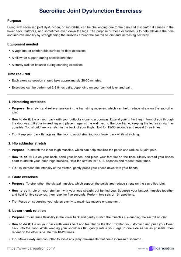
Purpose
Knee X-rays are pivotal for visualizing the knee joint and its surrounding bone structures, aiding healthcare providers in diagnosing conditions such as acute knee injuries, fractures, arthritis, and dislocations. Similarly, knee radiographs are critical in identifying conditions like anterior cruciate ligament injuries in sports and accidents, which frequently occur in the right knee. This imaging technique is indispensable for evaluating the knee in cases of injury or persistent pain and monitoring the progression of knee disorders over time.
Standard projections
Standard projections in knee radiography are essential for providing clear images of the knee’s structure from different angles. Here are the most commonly used views in lateral radiographs in clinical practice:
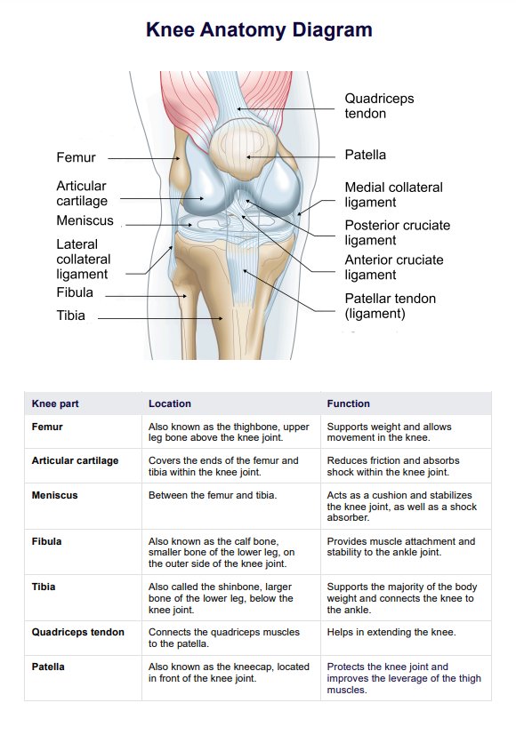
- Anteroposterior (AP) view: This standard projection delivers a frontal image of the knee, making it particularly effective for examining the overall alignment, detecting fractures, and evaluating the joint spaces. It is vital for assessing the general health of the knee joint and the surrounding bones. Additionally, it offers an excellent view of the medial collateral ligament, enabling a thorough assessment of its integrity and any related injuries.
- Horizontal beam lateral view: This lateral projection requires the patient to lie down, with the X-ray beam passing horizontally through the knee. It is essential for capturing the knee joint’s side profile, facilitating a detailed examination of the joint spaces, and detecting potential joint effusions or fluid accumulation. The horizontal beam lateral view is specifically designed to evaluate the lateral collateral ligament and other structures on the outer side of the knee. Moreover, this view is crucial for identifying avulsion fractures, clearly depicting bone fragments and their alignment.
Other projections
Additional projections in knee radiography are used to provide different perspectives and detailed views of specific areas within the knee joint. Each projection serves a unique purpose in the diagnostic process:
- Rolled lateral view: This projection offers a more distinct view of the femoral condyles and the tibial plateau, with no superimposition of the fibula over the tibia. It is especially valuable for identifying subtle fractures and providing a detailed assessment of the joint surfaces, including a clear image of the lateral femoral condyle. A lateral knee radiograph is crucial for a thorough evaluation, enabling clinicians to examine the knee from multiple perspectives for comprehensive diagnostic insights.
- Knee AP weight-bearing view: Conducted while the patient stands, this view replicates the natural load and stress experienced by the knee during standing. It yields critical insights into the knee’s functional alignment and is essential for diagnosing conditions such as osteoarthritis. Additionally, this view is particularly effective for evaluating the impact of varus stress on the knee’s alignment and structural integrity, providing vital information for targeted treatment strategies.
- Rosenberg’s view: This view is tailored specifically to examine the posterior aspects of the knee joint. Performed with the patient’s knees bent at a 45-degree angle while bearing weight, it provides an enhanced view of the joint space, making it particularly valuable for diagnosing or monitoring the progression of osteoarthritis. Additionally, this view is especially effective for evaluating the posterior cruciate ligament.
- Posteroanterior flexion weight-bearing view (PA Flex): This projection is similar to Rosenberg’s view but with the patient flexing their knees at about 30 degrees. It helps in evaluating joint space narrowing and other changes linked to osteoarthritis.
- Skyline or merchant view: This projection is performed with the patient either lying or sitting with their knees flexed, and the X-ray beam is directed downwards at the patella (kneecap). It is excellent for assessing the patellofemoral joint and detecting issues like patellar tracking abnormalities or chondromalacia patellae.
Preparations for knee radiographs
Preparation for a knee radiograph typically involves removing clothing, jewelry, or other objects that might obscure the X-ray images. Patients are often provided with a gown to wear. They may be asked to adopt specific positions to get clear views of the patient’s knee and structure.
In some cases, contrast material may be used to enhance the visibility of certain internal structures on the X-ray. The radiology technologist usually gives instructions to ensure the best possible image quality while minimizing exposure to radiation.
Knee Radiograph Results Template
Knee Radiograph Results Template Example
How to use our Knee Radiograph Results Template
Our free Knee Radiograph Results Template is free and easy to use. Here's how it's laid out:
Organization of the template
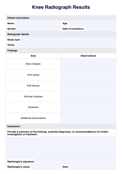
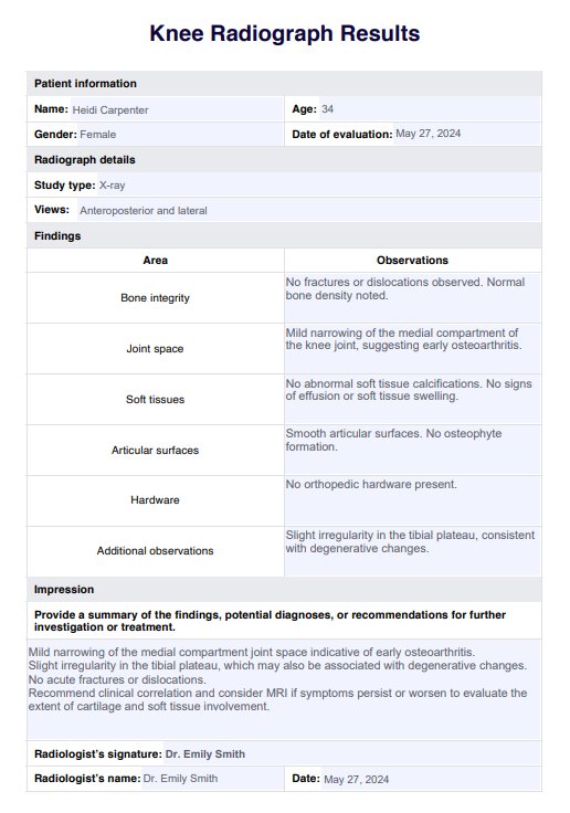
The template for knee radiograph results is designed to capture all relevant information clearly and organized. The Patient Information section includes fields for essential personal and clinical details such as the patient's name, age, sex, date of the exam, and referring physician. This ensures that the radiograph can be correctly linked to the patient’s medical records. The Radiograph Details section specifies the type of study and the views taken, which are crucial for understanding the scope of the examination.
The Findings section is structured to facilitate a detailed examination of various aspects of the knee. This includes bone integrity, joint space, soft tissues, articular surfaces, and any present hardware. The template ensures that each aspect is thoroughly assessed and documented, which is vital for a comprehensive evaluation. An Additional Observations field is included for any findings that don’t fit into the standard categories, allowing for flexibility in reporting.
Presenting and discussing results
Using the template effectively involves a detailed and systematic approach to documenting the radiographic findings. Each section is designed to capture specific types of information, which helps maintain clarity and focus throughout the report.
For instance, in the Findings section, the radiologist is encouraged to describe each observed condition, ensuring nothing is overlooked. The Impression section then allows for a summary of these findings, where the radiologist can suggest potential diagnoses or recommend further investigations.
This structured approach facilitates clear communication among medical professionals and enhances the consistency and accuracy of the reports. By providing a complete and precise examination record, the template assists healthcare providers in making informed decisions regarding patient management. Moreover, the standardized format supports effective collaboration and discussion among different medical teams, which is essential for optimal patient care.
Benefits of using this results template
Using a structured results template to present and discuss information significantly enhances clarity and readability. Organizing information with clear headings and subheadings makes it easier for readers to follow the narrative and grasp the main points. This structured approach helps identify different sections and their details quickly and improves the overall user experience by reducing confusion.
Additionally, a well-organized template aids in retaining information. When data is presented logically, it creates a clear mental map, making it easier for readers to remember the information. This is particularly beneficial in educational settings where understanding and recalling complex data is essential.
Moreover, this format allows for efficient information retrieval, which is crucial in both professional and academic environments. Users can quickly access specific data or insights without sifting through unstructured content.
Using a standardized template also ensures consistency in communication, which is important across various documents or presentations. It covers all necessary points uniformly, maintaining a consistent level of understanding and preventing discrepancies in information sharing.
Treatments for knee joint pathologies
Treatments for various knee injuries and joint pathologies vary widely depending on the specific condition, its severity, and the patient’s overall health. Effective management may involve a combination of non-surgical and surgical approaches tailored to address each individual’s unique needs. Specific radiographic views highlighting the posterior knee structure facilitate accurately diagnosing posterior cruciate ligament tears.
Physical therapy
Physical therapy is often the first line of treatment and is crucial for improving joint function, strengthening the muscles around the knee, and enhancing flexibility. Techniques such as exercises, stretching, and manual therapy are employed to reduce pain and increase mobility in the normal knee.
Medications
Medications are effective for pain management and reducing inflammation:
- Pain relievers and anti-inflammatory drugs: Over-the-counter medications like ibuprofen and acetaminophen can help reduce pain and inflammation. For more severe cases, stronger prescription medications may be necessary.
- Corticosteroids: Injections of corticosteroids directly into the knee joint can provide rapid relief of inflammation and pain. However, the effects are temporary, and repeated use can lead to joint damage over time.
Injections
Injections are useful for providing direct relief and promoting healing:
- Hyaluronic acid supplements: These injections are often used for treating osteoarthritis by providing lubrication to the knee joint, which can improve movement and reduce pain.
- Platelet-rich plasma (PRP): PRP injections use a concentration of a patient’s own platelets to promote the healing of injured tendons, ligaments, muscles, and joints.
Assistive devices
Braces and other devices can stabilize and protect the left knee:
- Braces or orthotics: For those with significant knee pain or instability, braces or orthotics can stabilize the knee and distribute forces away from the damaged part of the joint.
- Walking aids: Walking aids like canes or walkers may also be recommended to help reduce stress on the knee.
Lifestyle modifications
Changes to reduce stress on the joints and improve quality of life:
- Weight loss: Reducing body weight to decrease stress on knee joints.
- Activity modification: Limiting certain activities that exacerbate pain.
Surgical options
When non-surgical treatments are insufficient, surgery may be considered:
- Arthroscopic surgery: This minimally invasive surgery can repair or clean out damaged cartilage or remove bone fragments from within the joint.
- Partial knee replacement: This surgery involves replacing only the most damaged parts of the knee, preserving as much of the natural knee as possible.
- Total knee replacement: In cases where the knee joint is severely damaged by arthritis or injury, replacing the entire joint with an artificial one may be the best option to restore function and relieve pain.
Each treatment option has its benefits and risks, and the best approach depends on individual factors such as the type of pathology, the patient’s age, activity level, and overall health. Consulting with healthcare professionals like orthopedists, physical therapists, and rheumatologists is essential to developing a comprehensive treatment plan tailored to the patient’s needs.
Commonly asked questions
Knee radiographs are typically performed to diagnose fractures, monitor the progression of degenerative diseases like arthritis, and evaluate the structural integrity of the knee following an injury.
Knee radiographs provide detailed images of the bone structure and alignment of the joint line of the knee, aiding doctors in planning appropriate treatments such as surgery, physical therapy, or medication adjustments. They are also instrumental in confirming an anterior cruciate ligament injury, guiding subsequent treatment options.
Knee radiographs involve a low level of ionizing radiation exposure; however, the risk is generally very low compared to the benefits of diagnosing and treating knee conditions effectively.
Related Templates

Popular Templates