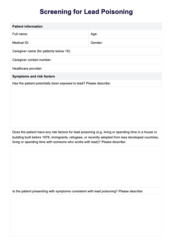
Ankle Radiograph Results Template
Explore our Ankle Radiograph Results Template for detailed, structured radiographic reporting and accurate diagnosis of ankle conditions.

Fact Checked by Ericka Pingol.


What is an ankle radiograph?
An ankle radiograph, commonly known as an ankle X-ray, is a diagnostic imaging technique used to visualize the bones and joints of the ankle. This test helps medical professionals assess the ankle for fractures, dislocations, arthritis, and other conditions. The radiograph uses a small amount of ionizing radiation to produce images of the ankle, which can show the alignment of the bones and the condition of the joint spaces and surrounding tissues.
Purpose
The primary purpose of an ankle radiograph is to evaluate the ankle following injury to diagnose fractures or dislocations. It is also used to monitor the progression of diseases such as osteoarthritis or rheumatoid arthritis.
Additionally, ankle radiographs can help plan medical or surgical treatments and assess healing after a fracture or surgery. The radiographs evaluate the condition of both the lateral and medial malleoli, helping diagnose fractures and track disease progression.
The three views of ankle radiographs
Ankle radiographs typically consist of three views, which provide different angles for thorough examination:
- Anteroposterior (AP): This is the frontal view of the ankle. The X-ray beam passes from the front to the back of the ankle. It helps assess the alignment of the tibia, fibula, talus, and joint spaces. The AP view is especially valuable for assessing the alignment of the distal tibia with the talus and the tibial articular surface.
- Mortise: This view is similar to the AP view, but the foot is rotated slightly. This view is particularly important for evaluating the ankle mortise, a U-shaped notch between the tibia and the fibula where the talus fits. It provides a clear image of the joint space and the integrity of the ankle mortise. The mortise view also provides a detailed examination of the ankle mortise, focusing on the alignment and space between the lateral malleolus and medial malleolus.
- Lateral: The X-ray beam passes from one side of the ankle to the other. This side view helps visualize the ankle's overlapping bones, the alignment of the tibia and fibula with the talus, and the posterior and anterior aspects of the ankle joint. The lateral view not only helps visualize the distal fibula but also provides insight into the condition of the proximal fibula.
These three views are crucial for a comprehensive assessment of the ankle, allowing for accurate diagnosis and appropriate management of ankle-related conditions.
Preparations for ankle radiographs
Several important steps must be taken when preparing for an ankle radiograph to ensure a smooth imaging process and accurate results. First, patients are asked to remove any clothing that covers the ankle area, along with all jewelry and metal objects that might obscure the ankle's view or interfere with the X-ray imaging. Sometimes, patients may be given a hospital gown to wear during the procedure.
Positioning for the X-ray is critical for capturing the best possible image. The radiologic technologist will guide the patient on positioning their ankle for each required view. For the anteroposterior view, the foot is placed flat and facing forward. The foot is slightly rotated inward for the mortise view, and for the lateral view, the patient is asked to turn the foot sideways.
Additionally, protective measures are often taken to minimize radiation exposure. Protective lead aprons or shields are used to cover areas of the body not being imaged, such as the pelvis, particularly in pregnant or reproductive-age patients.
It is also crucial for the patient to remain still during the X-ray to prevent the images from blurring. The technologist will help the patient find a comfortable yet firm position that can be maintained for a few seconds while the X-ray is taken. These steps are essential for ensuring that the ankle radiograph is clear and detailed, providing valuable assistance in accurate diagnosis and treatment planning.
How do professionals convey ankle radiograph results?
Once an ankle radiograph is taken, a radiologist specializing in interpreting medical images will review the X-rays. If available, they will look for any signs of injury, disease, or abnormality in the ankle and compare them with previous images. Part of the assessment includes examining the talar dome for smoothness and integrity, which is crucial for ankle mobility and stability.
The radiologist will then write a detailed report outlining their findings, noting any changes and potential concerns. The radiologist also assesses for any signs of ligamentous injury, which are crucial for determining the full extent of ankle trauma. They also look at the integrity of the deltoid ligament, particularly its lateral aspect, for any signs of injury or strain.
The report is sent to the physician who requested the X-ray. Depending on the context of the examination, this might be the patient’s general practitioner, an emergency room physician, or an orthopedic specialist. The referring physician will review the radiologist's report and may consult with the radiologist for further clarification or discussion about the findings.
The final step involves the physician discussing the results with the patient. During this consultation, the physician explains the radiograph findings, what they mean for the patient's health, and the next steps in terms of treatment or further testing. This discussion is crucial as it helps the patient understand their medical condition and the implications of the radiograph results, ensuring they are fully informed about their health and care options.
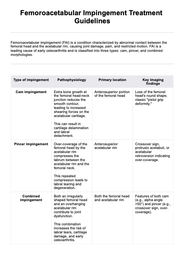
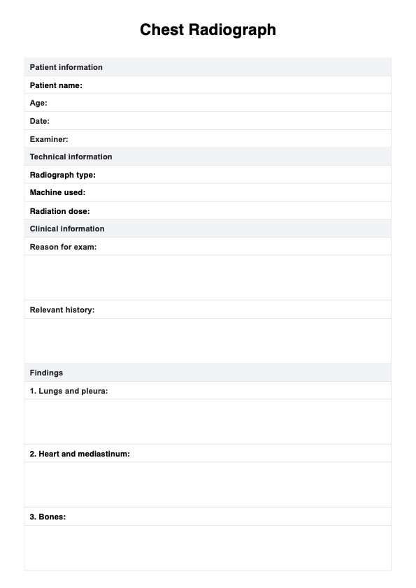
Ankle Radiograph Results Template
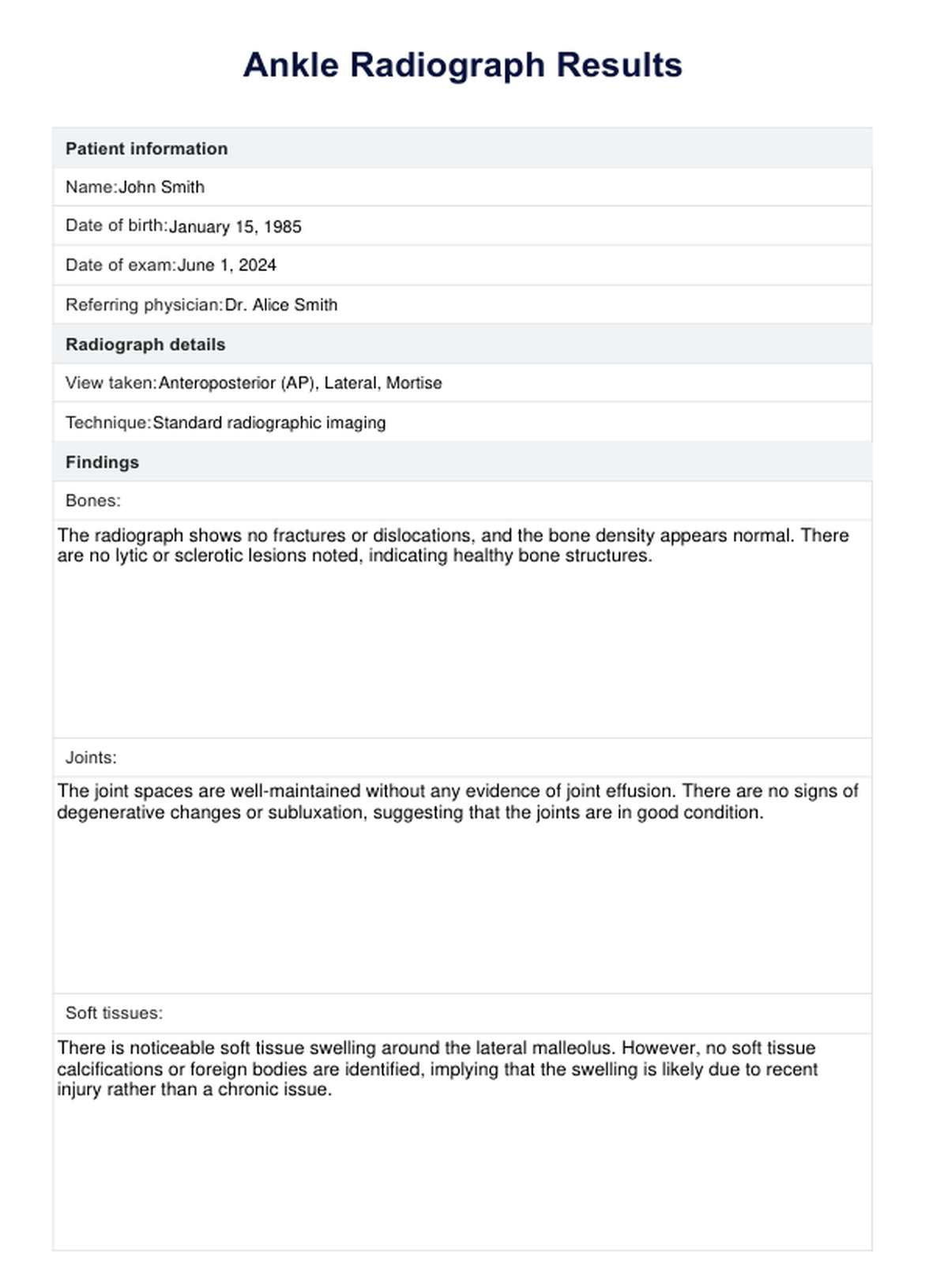
Ankle Radiograph Results Template Example
How to use our Ankle Radiograph Results Template
The Ankle Radiograph Results Template streamlines the documentation of radiographic findings. It ensures all necessary details are captured clearly and systematically, aiding radiologists in presenting consistent, interpretable results to referring physicians. Here's how to effectively use our template:
Step 1: Fill in patient information
The template starts by gathering essential identifiers for the patient. This includes the patient's name, date of birth, exam date, and referring physician. Accurate entry of this information ensures the radiograph is correctly associated with the patient's medical records.
Step 2: Note radiograph details
This section records specifics about the radiographic technique and the views taken. Documenting these details is critical as it provides context for the images and helps interpret the findings accurately based on how the radiograph was captured.
Step 3: Enter findings in the table
The results are organized into a tabular format, which is divided into categories—bones, joints, and soft tissues. This structure allows for:
- Bones: Detailed entries about bone integrity, fractures, and other abnormalities.
- Joints: Observations on joint alignment and signs of diseases such as arthritis.
- Soft tissues: Notes on the condition of surrounding soft tissues, including any swelling or irregularities.
Step 4: Add additional observations
This section is reserved for any significant findings that do not fit into the standard categories but are essential for a comprehensive diagnostic overview.
Step 5: Summarize the impression
Here, the radiologist summarizes the radiographic findings, offering an overall impression or diagnosis. This critical analysis guides the referring physician’s next steps in patient management.
Benefits of using this results template
Using a standardized results template enhances clarity and consistency in presenting information. It allows for easy comparison between different datasets or studies. This uniform approach facilitates understanding among diverse audiences, including stakeholders and decision-makers.
A results template also streamlines the review process and helps digest complex data. By providing a predefined format for data entry and reporting, it promotes accuracy and reduces errors. This method supports transparency and efficiency, especially in collaborative or multidisciplinary projects where consistent communication is crucial.
Treatments for ankle fractures
Ankle fractures are treated based on their severity, location, and the patient's health and activity level. Treatment approaches also vary depending on the specific fracture, whether it's a medial malleolus fracture or a lateral malleolus fracture. Specific fractures like a talar neck fracture require precise imaging for accurate diagnosis and targeted treatment planning.
Initial treatment often includes the RICE protocol—rest, ice, compression, and elevation—which reduces swelling and pain. For less severe fractures, a cast or splint may be used to immobilize the ankle, helping the bones heal properly. Physical therapy is commonly recommended to restore range of motion, strength, and balance.
For more complex fractures, surgical intervention may be necessary. Surgery typically involves using plates, screws, or rods to stabilize the bones and ensure proper alignment. After surgery, the ankle is usually immobilized with a cast or boot for several weeks.
Rehabilitation follows surgical treatment to help the patient regain full function. The specific approach to treating an ankle fracture depends on the precise nature of the fracture and the individual's needs, emphasizing a personalized treatment plan for optimal recovery.
Commonly asked questions
An ankle radiograph, or X-ray, is primarily used to detect fractures, assess joint alignment, and identify signs of arthritis or other bone conditions in the ankle.
Generally, no special preparation is required for an ankle radiograph. You may be asked to remove jewelry or any metal objects that could interfere with the image clarity.
Ankle radiographs involve a very low dose of radiation, making them safe for most people. However, pregnant women should inform their doctor as radiation can pose risks to the developing fetus.
Related Templates

Popular Templates