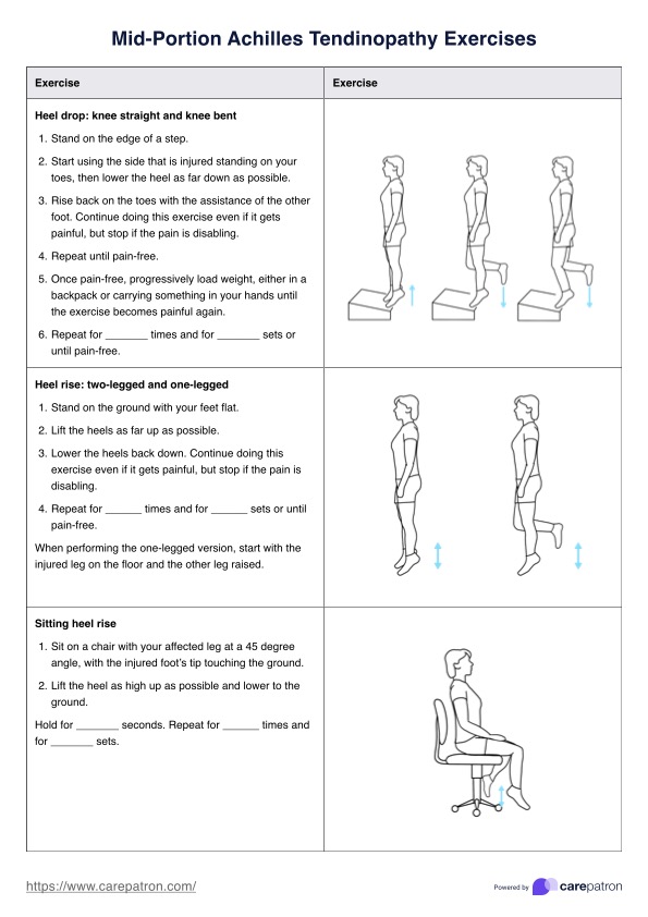
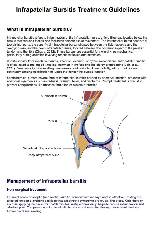
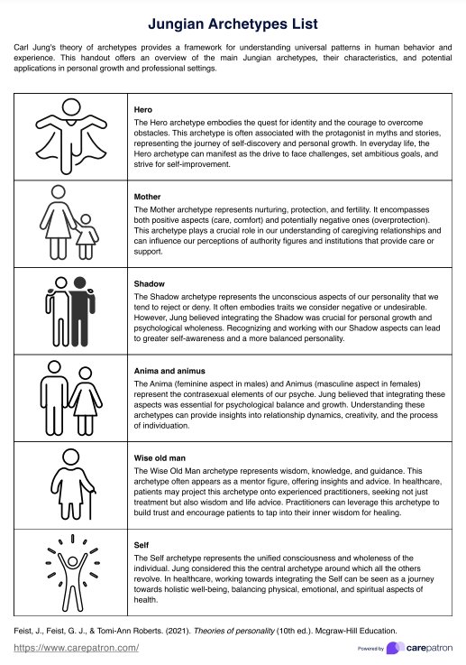
Ulnar Nerve Anatomy Diagram
Read our ulnar nerve anatomy guide for healthcare professionals. Download our free Ulnar Nerve Anatomy Diagram template for educational use here.

Fact Checked by Ericka Pingol.


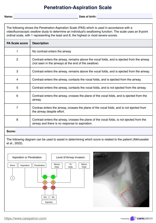
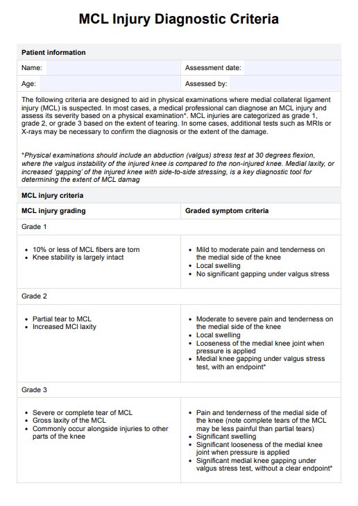
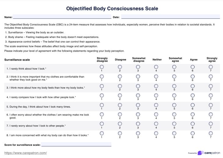
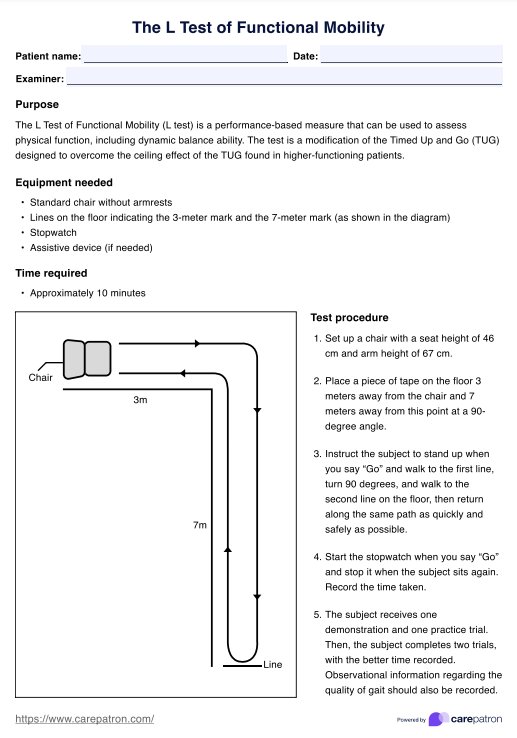
Ulnar nerve pathway
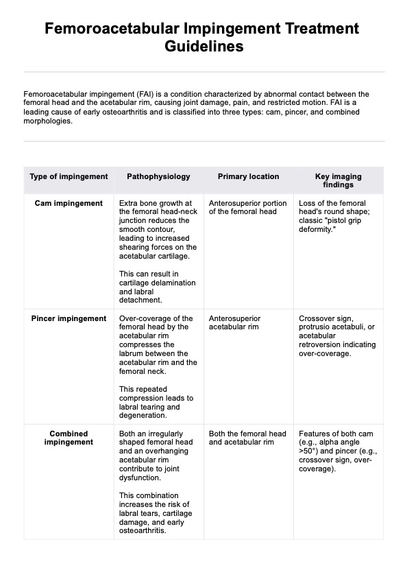
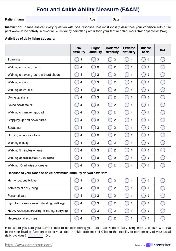
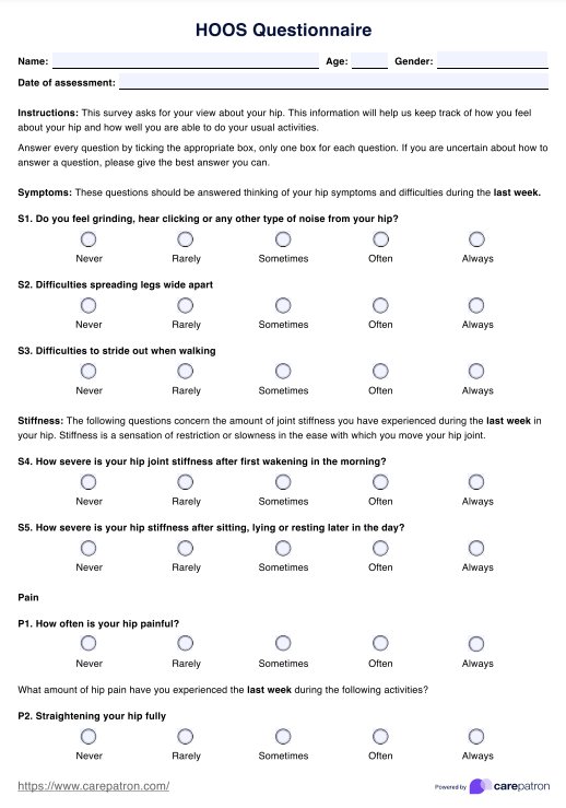
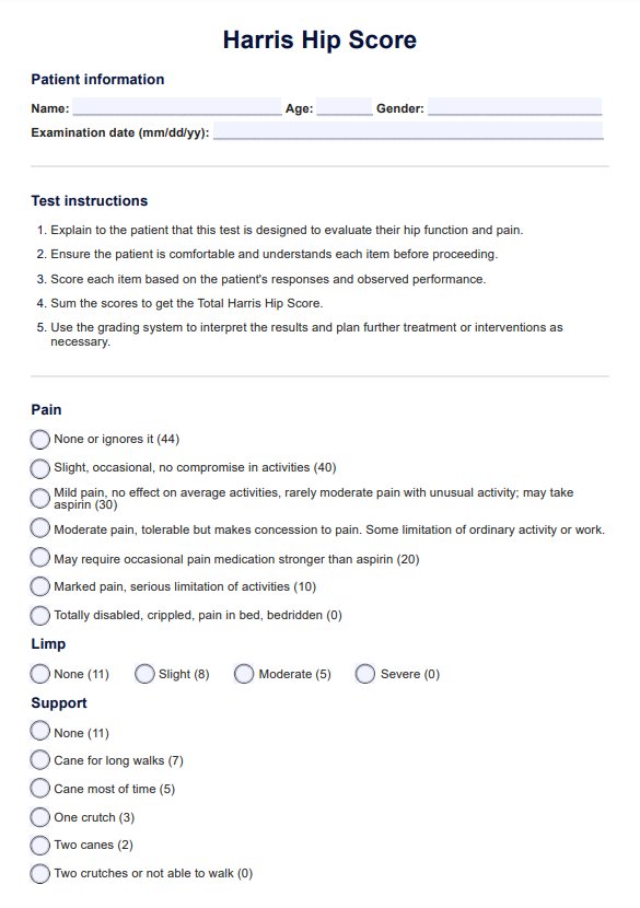
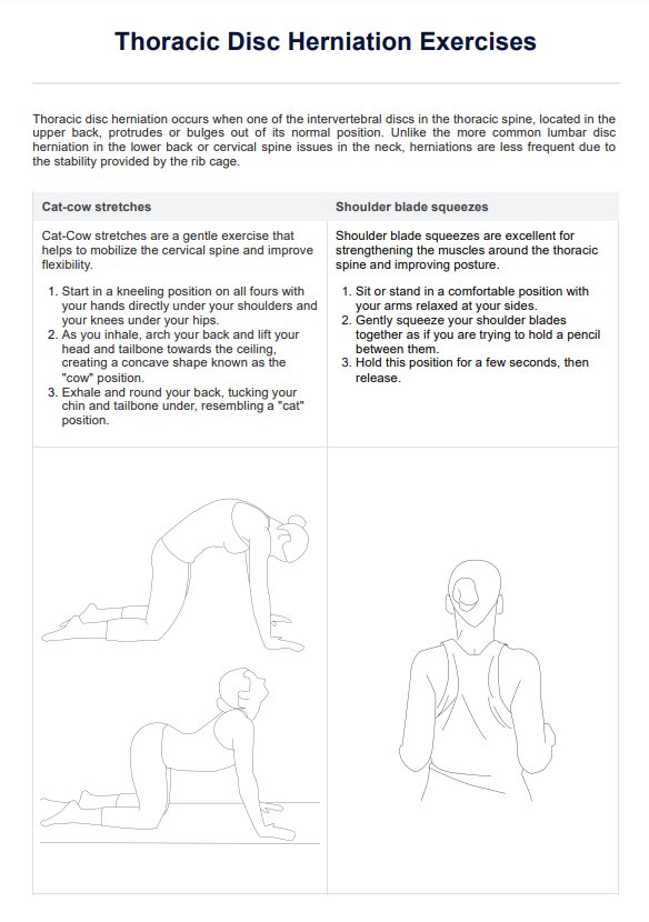
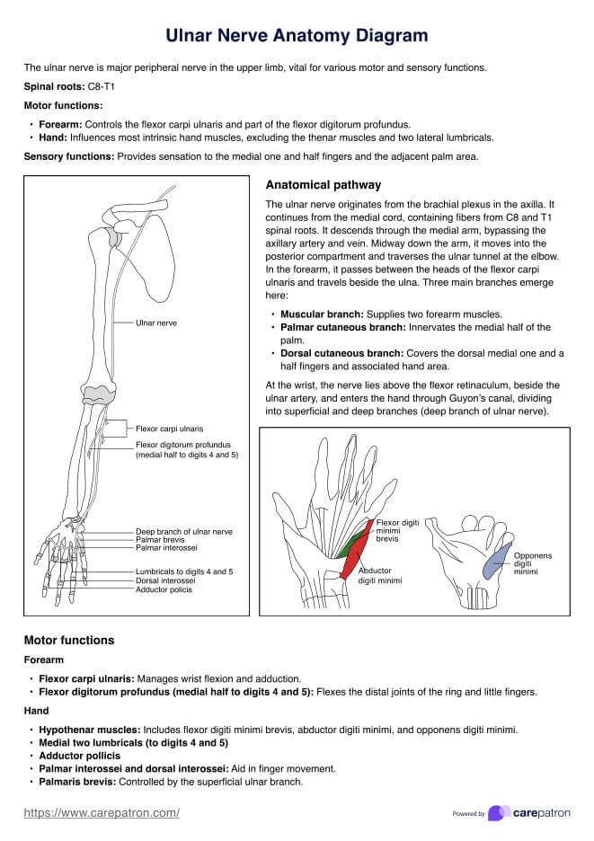
The ulnar nerve, a major peripheral nerve of the upper limb, is crucial in motor and sensory functions. Originating from the C8 and T1 spinal nerve roots, it is a continuation of the medial cord of the brachial plexus. This nerve provides functionality and sensation to muscles in the forearm and hand.
The ulnar nerve originates from the axilla (armpit) region as it exits the brachial plexus, running down the medial aspect of the upper arm, alongside the brachial artery. At the midpoint of the arm, ulnar nerve passes through the medial fascial septum, entering the posterior compartment. This pathway continues as the nerve passes posteriorly to the medial head, traversing through the cubital tunnel—a space between the medial epicondyle of the humerus and the olecranon of the ulna.
In the forearm, the ulnar nerve pierces the two heads of the flexor carpi ulnaris muscle and travels deep along the ulna, where three significant branches arise:
- Muscular branch: Innervates the flexor carpi ulnaris and the medial half of the flexor digitorum profundus.
- Palmar cutaneous branch: Provides sensation to the medial half of the palm.
- Dorsal cutaneous branch: Supplies the dorsal surface of the medial one and a half fingers and associated hand area.
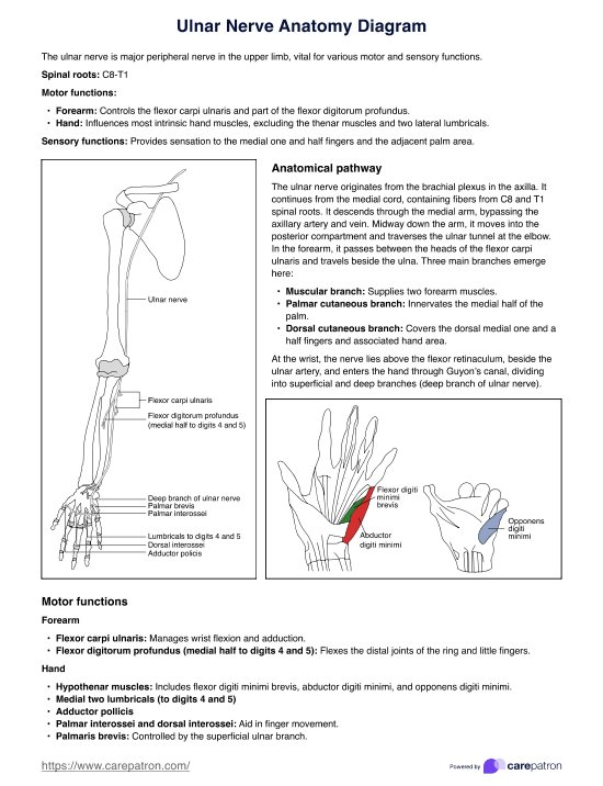
At the wrist, the ulnar nerve lies superficial to the flexor retinaculum, adjacent to the ulnar artery. It enters the hand through Guyon’s canal, where it divides into superficial and deep branches. The superficial branch innervates the palmaris brevis muscle and provides sensation to the medial one and a half fingers. The deep branch supplies most of the intrinsic hand muscles, including the hypothenar muscles, the medial two lumbricals, the adductor pollicis, and the interossei muscles (both palmar and dorsal).
Understanding the pathway of the ulnar nerve is essential for diagnosing and treating conditions related to nerve damage, such as ulnar nerve palsy and cubital tunnel syndrome. Additionally, it is important to differentiate the ulnar nerve from the median nerve, which innervates the remaining muscles in the anterior forearm and lateral hand.
Download our free Ulnar Nerve Anatomy Diagram example here
Ulnar Nerve Anatomy Diagram Template
Ulnar Nerve Anatomy Diagram Sample
Commonly asked questions
The ulnar nerve is most commonly damaged at the elbow, particularly in the cubital tunnel, and at the wrist, often within Guyon’s canal.
The ulnar nerve arises from the C8 and T1 spinal nerve roots.
Symptoms of ulnar nerve injury include numbness or tingling in the ring and little fingers, weakness in hand grip, and difficulty with finger coordination.
Related Templates

Popular Templates