Syringomyelia Treatment Guidelines
Discover comprehensive syringomyelia treatment guidelines for healthcare professionals. Learn about diagnosis, treatment options, and management.


What is syringomyelia?
Syringomyelia is a chronic condition where a fluid-filled cyst, known as a syrinx, forms within the spinal cord. This cyst can expand over time, causing significant damage to the spinal cord and disrupting cerebrospinal fluid (CSF) flow. The condition is often associated with various factors, including brain and spinal cord malformations like Chiari malformation, spinal cord tumors, tethered spinal cord, and spinal cord injury.
In some cases, the cause remains unknown, leading to idiopathic syringomyelia. Magnetic resonance imaging (MRI) is the primary tool to diagnose this condition, revealing the syrinx and any associated abnormalities within the spinal canal. Left untreated, syringomyelia can result in further spinal cord injury, leading to severe neurological deficits.
Symptoms of syringomyelia
Syringomyelia presents with various symptoms that can progressively worsen if not treated. These symptoms are primarily due to the disruption of normal functions within the spinal cord.
- Chronic pain in the neck, shoulders, and back
- Weakness and stiffness in the arms and legs
- Loss of sensation, especially to temperature and pain
- Muscle atrophy and weakness
- Abnormal curvature of the spine (scoliosis)
- Headaches, particularly with straining
- Bladder and bowel dysfunction
Causes of syringomyelia
The development of syringomyelia is linked to several underlying conditions and factors. Understanding these causes is crucial for effective diagnosis and treatment.
- Chiari malformation: Brain tissue extends into the spinal canal, disrupting CSF flow.
- Spinal cord injury: Trauma can lead to the formation of a syrinx within the spinal cord.
- Spinal cord tumors: Growths can obstruct the flow of cerebrospinal fluid.
- Tethered spinal cord: A condition where the spinal cord is abnormally attached within the spinal canal, affecting CSF flow.
- Infections: Conditions like meningitis can cause inflammation and syrinx formation.
- Idiopathic syringomyelia: Cases where the exact cause is unknown.
- Congenital conditions: Birth defects involving the brain and spinal cord.
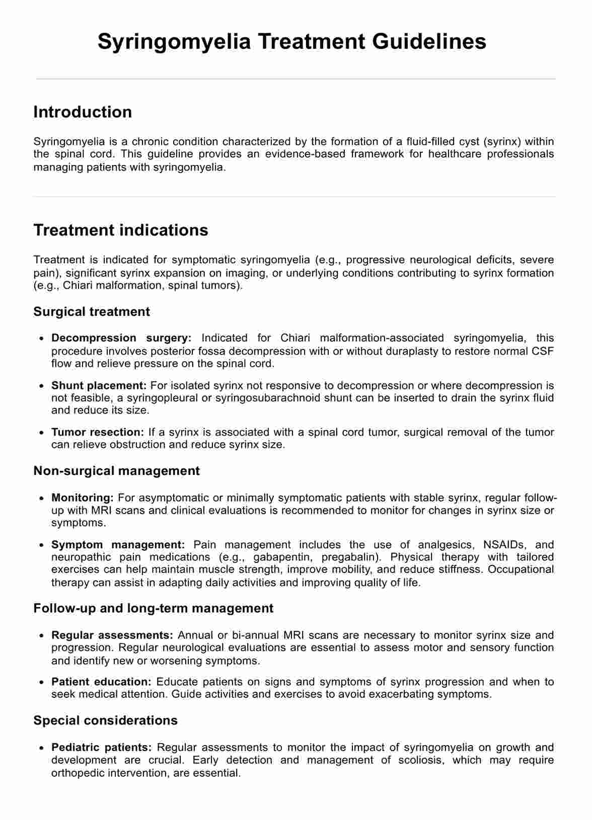
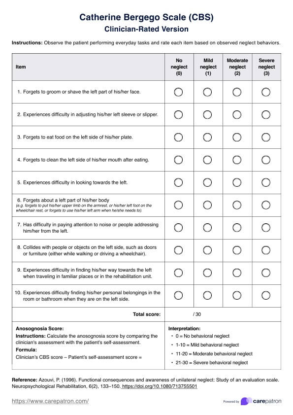
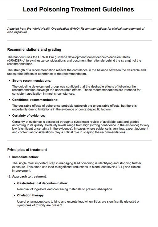
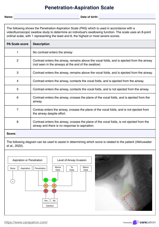
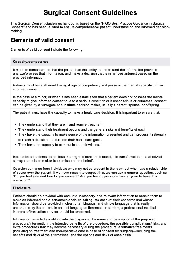
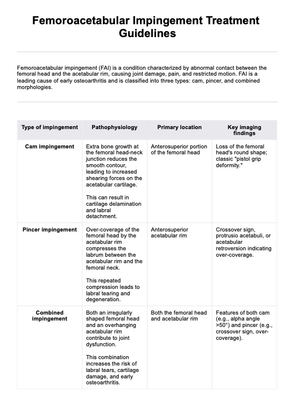
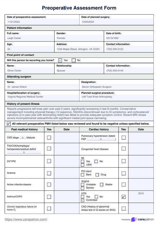
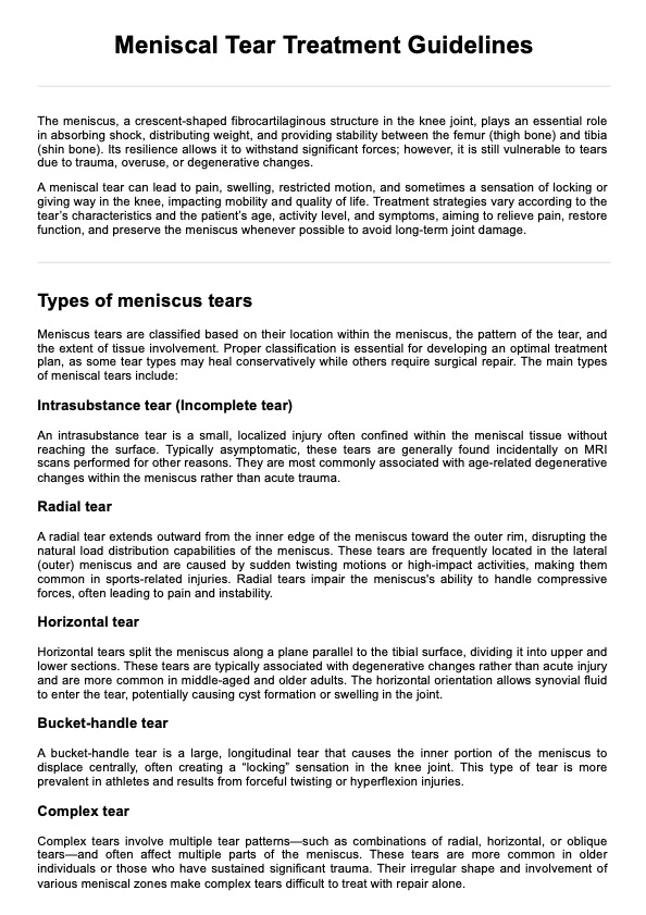
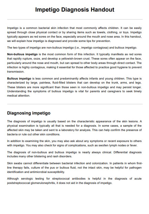
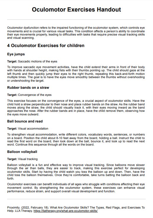
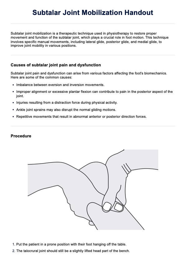
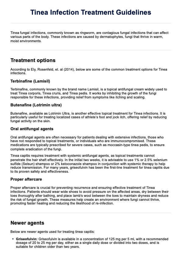
Syringomyelia Treatment Guidelines Template
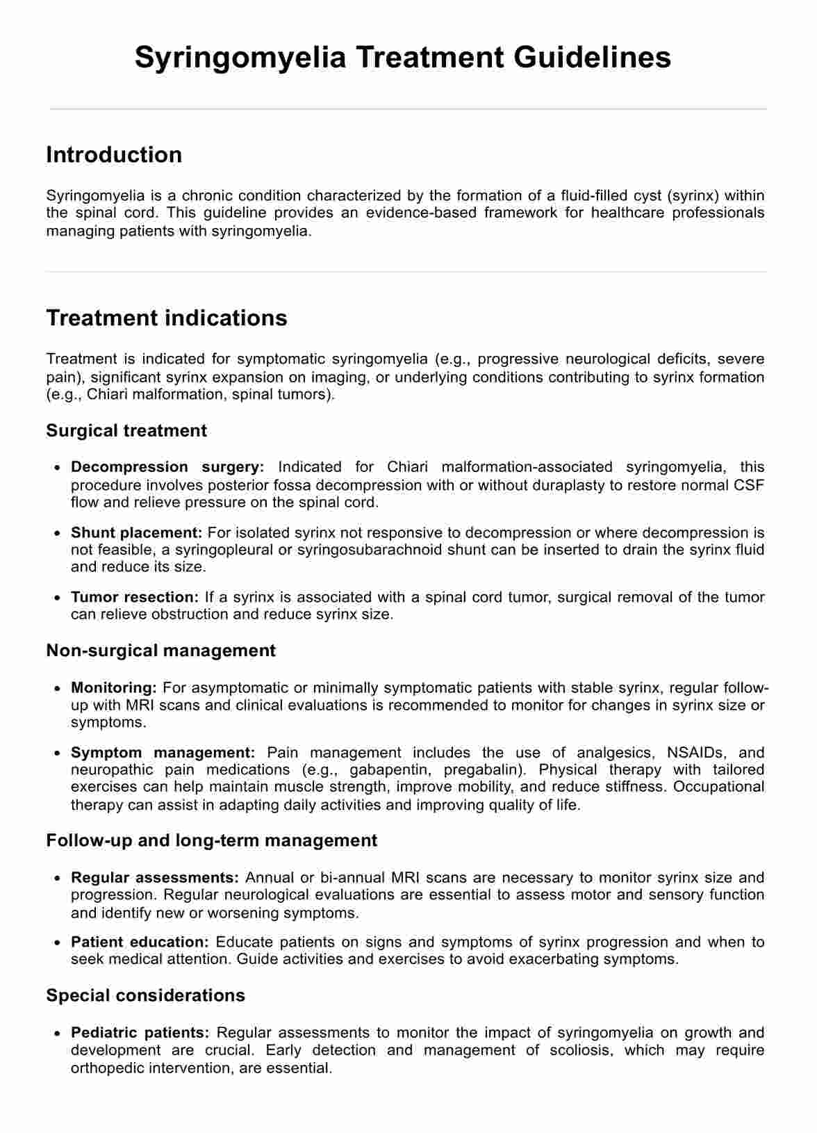
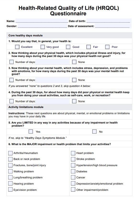
Syringomyelia Treatment Guidelines Example
How is syringomyelia diagnosed?
Diagnosing syringomyelia involves clinical evaluation and imaging techniques to identify a fluid-filled cyst within the spinal cord. The process begins with a medical history and physical examination to assess symptoms like muscle weakness and neurological function.
Magnetic resonance imaging (MRI) is the primary tool used to diagnose syringomyelia, revealing the cyst and associated abnormalities such as Chiari malformations, where brain tissue extends into the spinal canal. This helps distinguish between primary spinal syringomyelia and secondary causes like spinal cord injuries or inflammation of the spinal cord membranes.
How do healthcare professionals treat syringomyelia?
Treating syringomyelia involves a combination of surgical and non-surgical approaches to manage symptoms and prevent further neurological damage. Treatment choice depends on the underlying cause and severity of the condition.
1. Decompression surgery
Posterior fossa decompression is often performed to treat syringomyelia associated with Chiari malformation. This surgery relieves pressure and restores the normal flow of cerebrospinal fluid around the brain and spinal cord.
2. Shunt placement
In cases where decompression is not sufficient or feasible, a shunt may be placed to drain the syrinx and reduce its size, alleviating pressure on the spinal cord.
3. Tumor resection
If a spinal cord tumor is causing the syrinx, removing the tumor can relieve the obstruction and improve the flow of cerebrospinal fluid.
4. Tethered cord release
For syringomyelia caused by a tethered spinal cord, surgical release of the tethered cord can help restore normal movement and reduce symptoms.
5. Symptom management and monitoring
Regular monitoring with MRI and clinical evaluations, combined with physical therapy and pain management, helps manage symptoms and track the progression of congenital syringomyelia and other forms.
Commonly asked questions
Magnetic Resonance Imaging (MRI) is the primary tool for diagnosing syringomyelia, as it reveals the presence of a fluid-filled cyst within the spinal cord and any associated abnormalities.
Common surgical treatments include posterior fossa decompression for Chiari malformations, shunt placement to drain the syrinx, and tumor resection if a spinal cord tumor is present.
Non-surgical management includes regular monitoring with MRI, physical therapy, pain management, and occupational therapy to maintain muscle strength and improve quality of life.


