Supine Roll Test
Learn how to conduct and interpret the Supine Roll Test for diagnosing HC-BPPV. Download our template with sample answers and improve patient care today!


What is lateral benign paroxysmal positional vertigo (BPPV)?
Lateral benign paroxysmal positional vertigo (BPPV) is a subtype of benign paroxysmal positional vertigo, a common vestibular disorder that causes brief episodes of vertigo related to changes in head position. BPPV occurs when otoconia (calcium carbonate crystals) dislodge from the utricle and enter one of the semicircular canals, typically the posterior canal.
In lateral BPPV, also known as horizontal canal BPPV (HC-BPPV), these dislodged crystals move into the horizontal (lateral) semicircular canal, causing vertigo when the head is turned to the side. This condition can significantly impact balance and quality of life but is generally treatable through specific repositioning maneuvers.
Symptoms of lateral benign paroxysmal positional vertigo
Lateral BPPV is characterized by symptoms that typically occur when the head is moved in specific ways, triggering episodes of vertigo.
- Sudden, intense episodes of dizziness triggered by head movements, particularly when turning the head to the side.
- Involuntary, rapid eye movements (nystagmus) often observed when the head is turned or during specific diagnostic maneuvers.
- Nausea and vomiting can accompany vertigo, especially in severe cases.
- Difficulty maintaining balance, particularly when walking or moving quickly.
- Dizziness that occurs when changing positions, such as getting out of bed or looking up.
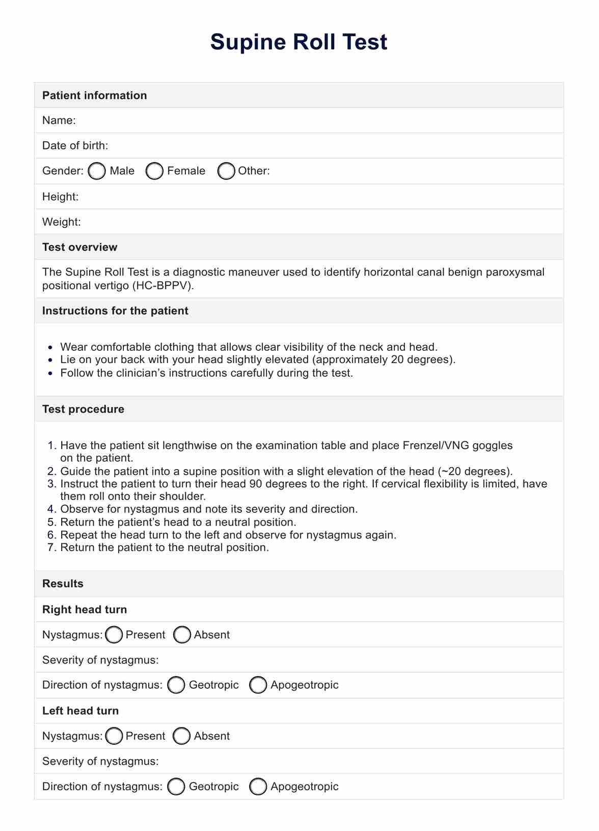
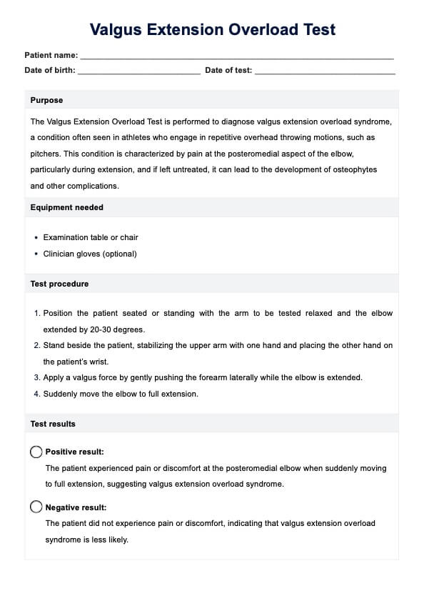
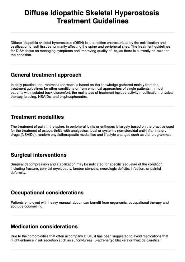
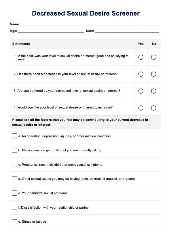
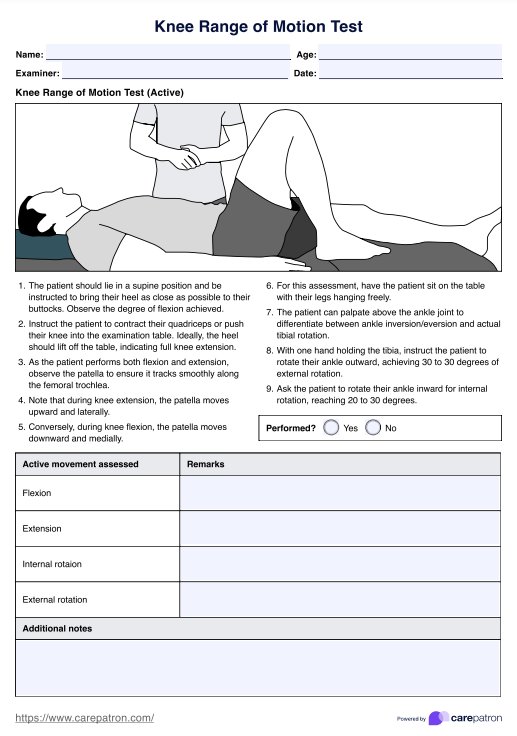
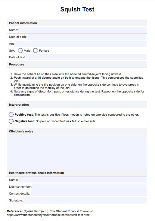
Supine Roll Test Template
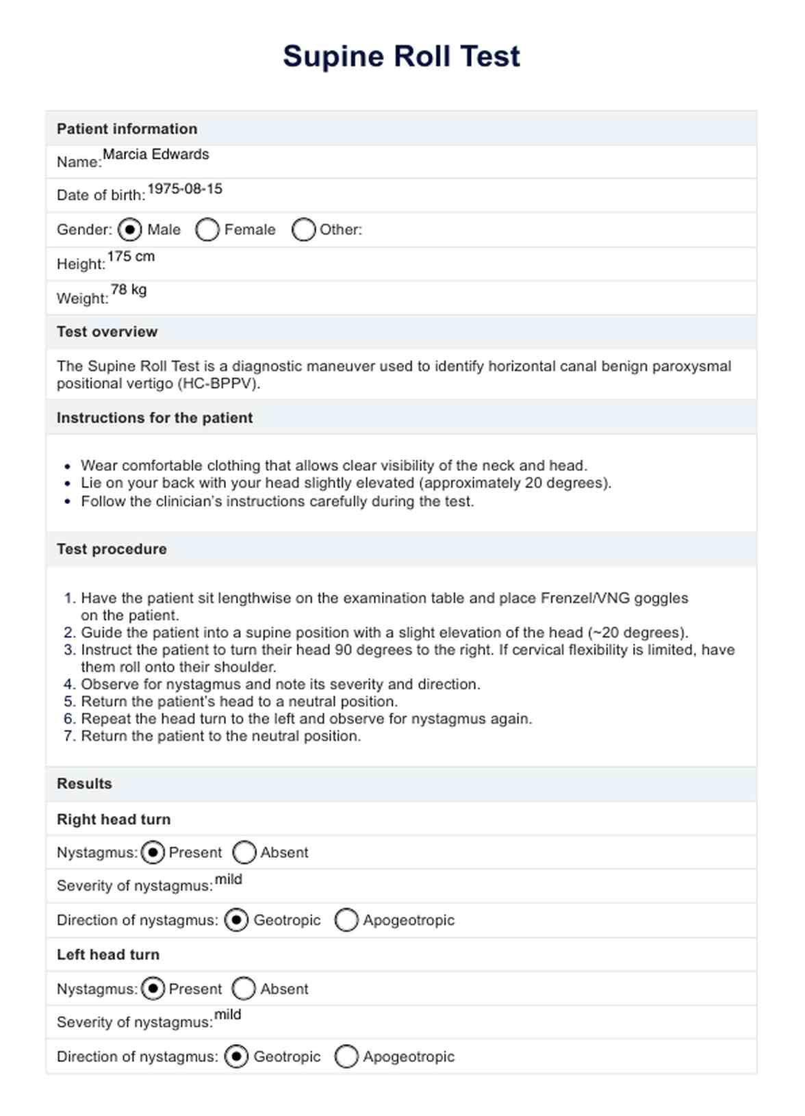
Supine Roll Test Example
Causes of lateral and horizontal canal BPPV
The primary cause of lateral BPPV is the dislodgement of otoconia from the utricle into the lateral semicircular canal. Several risk factors also can contribute to this occurrence.
- The primary cause is the dislodgement of otoconia from the utricle into the lateral semicircular canal. These particles disrupt the normal fluid flow in the canal, leading to abnormal signals being sent to the brain about head movement.
- Trauma to the head can cause the otoconia to dislodge and migrate into the semicircular canals.
- As people age, the otolithic membrane, where the otoconia are normally embedded, can degenerate, making the otoconia more likely to dislodge.
- Conditions such as labyrinthitis or vestibular neuritis can increase the risk of otoconia dislodgement.
- Extended periods of immobility or bed rest can contribute to the development of BPPV due to the lack of normal head movements.
What is the Supine Roll Test in the supine position?
The Supine Roll Test is a diagnostic maneuver used to identify horizontal canal benign paroxysmal positional vertigo. This condition is a subtype of benign paroxysmal positional vertigo, a common vestibular disorder that causes episodes of vertigo triggered by changes in head position.
In addition, the Supine Roll Test helps to determine if the dislodged otoconia (calcium carbonate crystals) are present in the horizontal (lateral) semicircular canal, leading to vertigo when the head is turned to the side. Another diagnostic maneuver used alongside the Supine Roll Test is the lean test, which assesses the characteristics of nystagmus induced by positional tests and can predict the recurrence and success of treatment for HC-BPPV.
How is this test conducted?
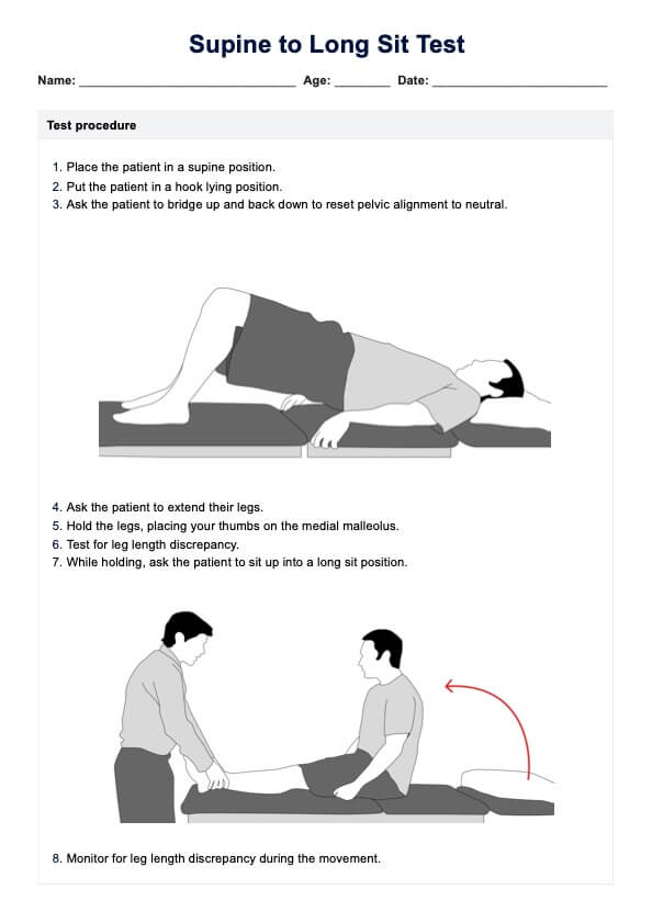
The Supine Roll Test is performed with the patient lying on their back (supine) on an examination table. The procedure involves several specific steps:
- The patient lies flat on their back with their head elevated slightly (approximately 20 degrees).
- The examiner turns the patient's head 90 degrees to one side while keeping the eyes open. The examiner keeps the patient's head in this position to observe any nystagmus (involuntary eye movements) and vertigo.
- The patient's head is then returned to the neutral, supine position.
- The examiner repeats the process by turning the patient's head 90 degrees to the opposite side, again observing for nystagmus and vertigo.
- Throughout the test, the examiner observes the patient's eyes for signs of nystagmus and inquires about any sensations of vertigo the patient may experience.
How are the results interpreted?
Interpreting the results of the Supine Roll Test involves assessing the presence and type of nystagmus and the patient's report of vertigo. The following interpretations are used:
- Geotropic nystagmus: If nystagmus beats towards the ground (geotropic), it suggests that the affected ear is on the side with the more intense nystagmus. This type of nystagmus indicates the presence of free-floating otoconia in the horizontal semicircular canal.
- Apogeotropic nystagmus: If nystagmus beats away from the ground (apogeotropic), it suggests that the affected ear is on the side with the less intense nystagmus. This type of nystagmus indicates that the otoconia are likely adhering to the canal wall.
- Severity and direction: The severity and direction of nystagmus provide clues about which side is affected and the extent of the condition. A positive test is confirmed by nystagmus and vertigo upon head turning.
The Supine Roll Test is an essential tool for diagnosing HC-BPPV and helps guide appropriate treatment strategies, such as repositioning maneuvers to move the otoconia out of the semicircular canal and alleviate symptoms.
Benefits of conducting this test
Conducting the Supine Roll Test offers several significant advantages for healthcare professionals diagnosing horizontal canal benign paroxysmal positional vertigo. These benefits ensure precise clinical diagnosis, effective patient management, and improved clinical outcomes.
The Supine Roll Test is particularly useful in assessing the vestibular system, as it helps identify the displacement of otoconia within the semicircular canals, which can lead to abnormal fluid displacement and trigger nystagmus, a key symptom of BPPV.
Accurate diagnosis
The Supine Roll Test provides a precise method for identifying HC-BPPV. By observing nystagmus (involuntary eye movements) and vertigo during head movements, healthcare professionals can accurately determine the affected inner ear and the specific type of BPPV. This accuracy is crucial for developing targeted treatment plans and ensuring effective patient care.
Non-invasive and practical
This non-invasive test involves simple positional maneuvers without requiring complex equipment or procedures. Its practicality allows for quick administration in various clinical settings, making it an accessible diagnostic tool. This ease of use ensures timely assessment and intervention, which is especially important in acute cases of vertigo where rapid relief is necessary.
Differentiation from other disorders
The Supine Roll Test helps differentiate HC-BPPV from other vestibular disorders with similar symptoms. Accurate differentiation ensures that patients receive appropriate treatments specific to their condition, reducing the risk of misdiagnosis. The clear, observable signs provided by nystagmus patterns enhance diagnostic accuracy and confidence in clinical decision-making.
Commonly asked questions
The primary purpose of the Supine Roll Test is to diagnose horizontal canal benign paroxysmal positional vertigo by identifying the presence and type of nystagmus when the patient's head is turned to the side.
A positive result is indicated by the presence of nystagmus and vertigo during the test. Geotropic nystagmus suggests free-floating otoconia in the horizontal canal, while apogeotropic nystagmus indicates otoconia adhering to the canal wall.
The Supine Roll Test is generally safe, but some patients may experience temporary discomfort or dizziness during the procedure. To ensure safety and accuracy, the test should be conducted by a trained healthcare professional.


