Plica Syndrome Test
Discover the comprehensive guide on Plica Syndrome, its diagnosis with the Mediopatellar Plica Test, and effective treatment options to enhance patient care.

Fact Checked by Ericka Pingol.


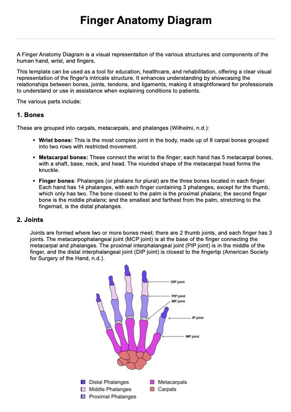
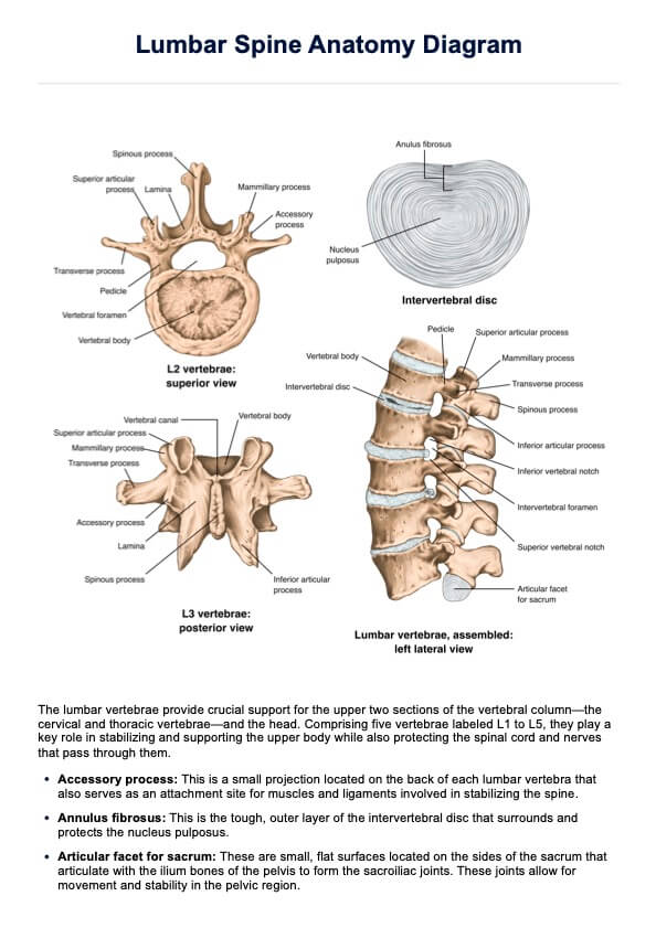
What is plica syndrome?
Plica syndrome occurs when a plica, a band of thick, fibrotic tissue within the knee, becomes inflamed or irritated. This condition often results from overuse or injury, causing friction between the plica and the patella or medial femoral condyle. Plica syndrome is characterized by anterior knee pain, which can significantly impact daily activities and athletic performance.
Symptoms of plica syndrome
Understanding the symptoms of plica syndrome is essential for early diagnosis and effective treatment. These symptoms can significantly impact daily activities and overall quality of life.
- Pain in the front of the knee
- Swelling or tenderness around the patella
- Clicking or snapping sensation when bending the knee
- Difficulty in fully extending the knee
- Increased pain with activities like squatting, running, or climbing stairs
Causes of plica syndrome
Several factors can contribute to the development of plica syndrome. Recognizing these causes can help prevent the condition and manage it effectively if it occurs.
- Overuse from repetitive activities such as running or cycling
- Knee trauma or injury
- Poor biomechanics or alignment issues
- Weakness or tightness in the muscles around the knee
- Inflammation due to conditions like rheumatoid arthritis
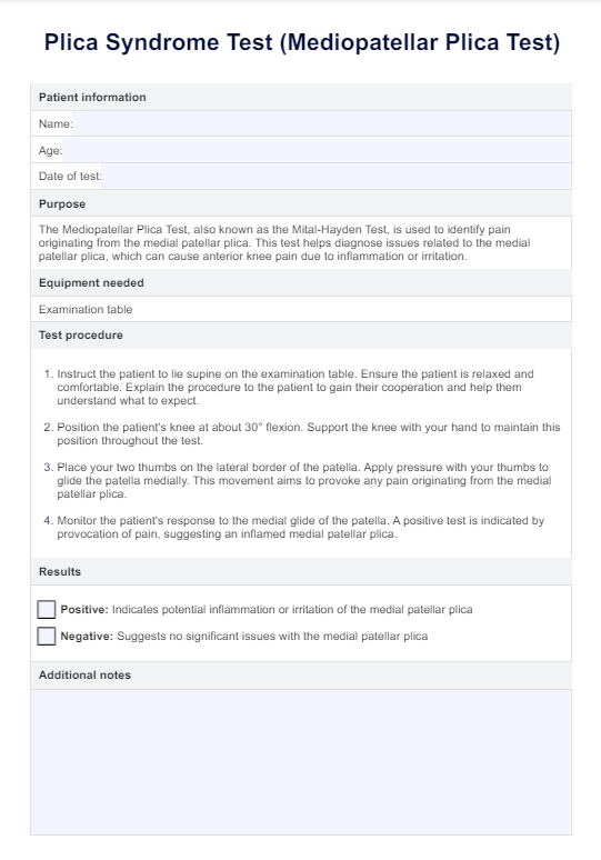
Plica Syndrome Test Template
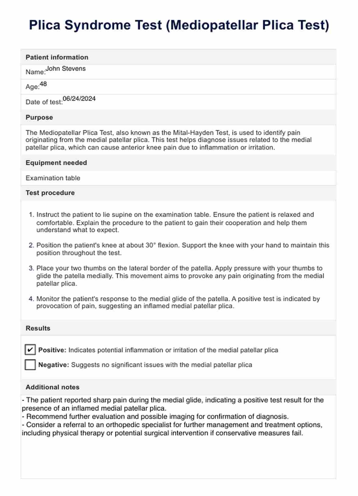
Plica Syndrome Test Example
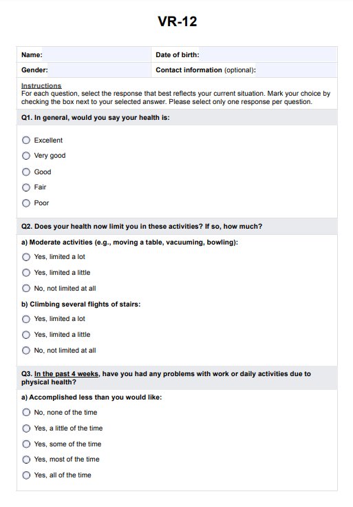
What is a mediopatellar plica test?
The mediopatellar plica test, also known as the Mital-Hayden Test, is used to identify pain originating from the medial patellar plica. This test is performed to diagnose plica syndrome, which can cause anterior knee pain due to the irritation of the synovial fold. By conducting this test, healthcare professionals can pinpoint the source of knee joint pain and determine if it is related to the medial patellar plica.
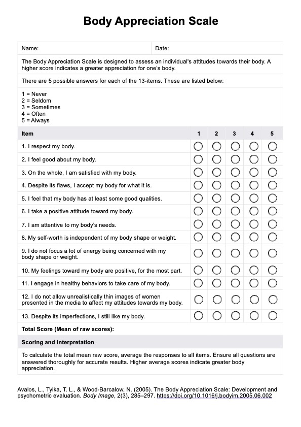
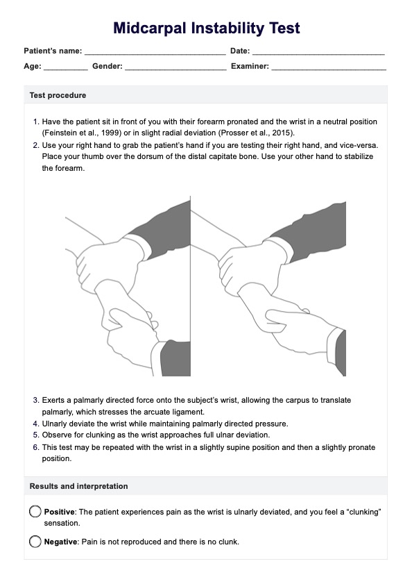
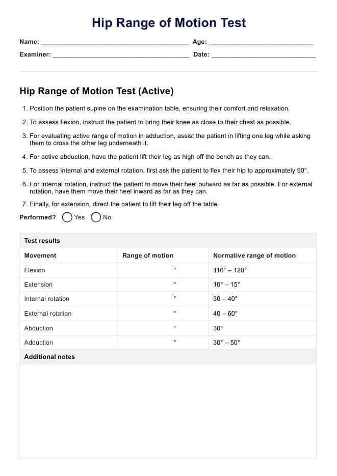
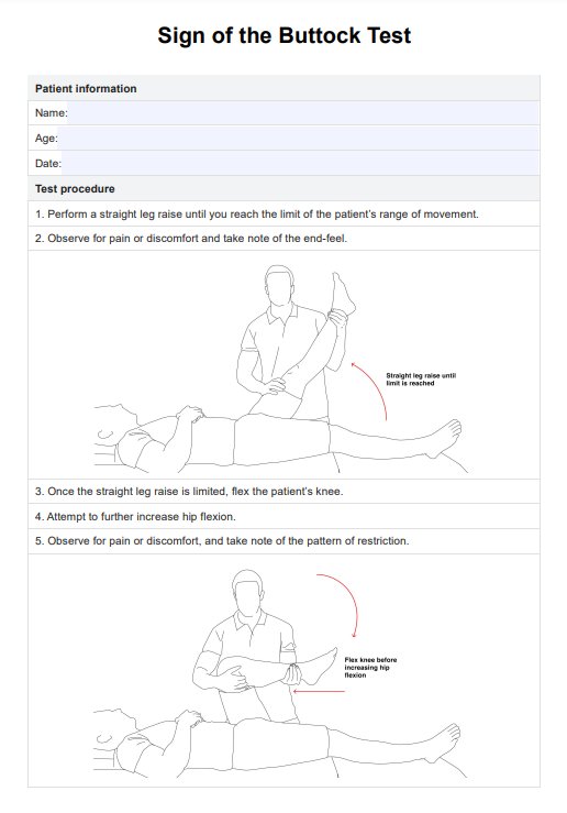
How is this test conducted?
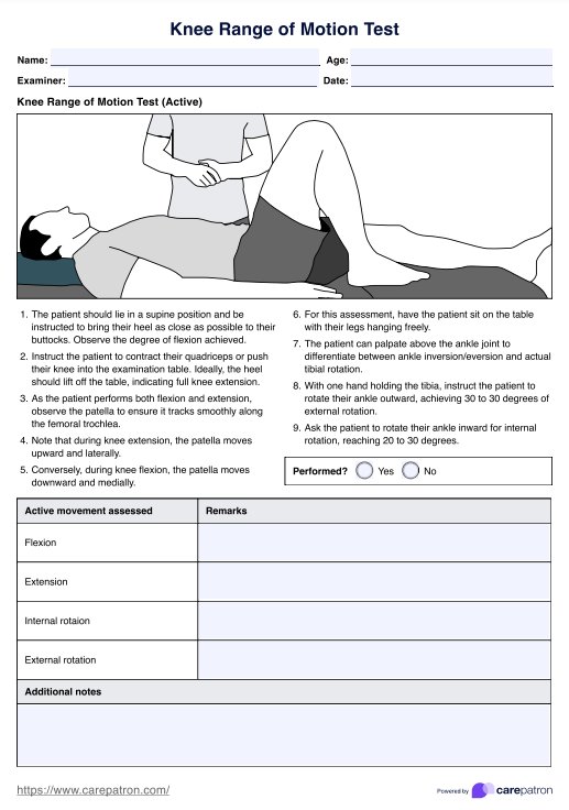
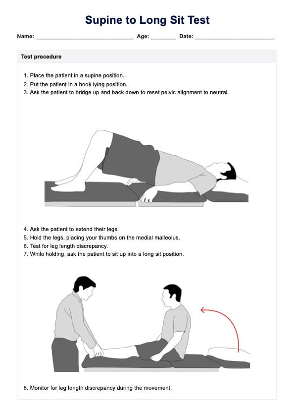
To conduct the mediopatellar plica test, the patient is positioned supine on an examination table with the knee flexed at about 30 degrees. The clinician supports the knee in this position. The clinician then places their thumbs on the lateral border of the medial patella plica and applies medial pressure to glide the patella inward. This maneuver aims to provoke any pain originating from the medial patellar plica.
The clinician closely monitors the patient's response to this pressure, looking for signs of pain or discomfort, indicating a positive result.
How are the results interpreted?
The results of the mediopatellar plica test are interpreted based on the patient's response to the patella's medial glide. A positive test is indicated by the provocation of pain during the medial glide, suggesting inflammation or irritation of the medial patellar plica. If the patient experiences significant pain or discomfort, it indicates the presence of plica syndrome.
A negative test, where no pain or discomfort is elicited, suggests that the medial patellar plica is not a significant source of the knee pain, and further investigation into other potential causes of knee problems may be necessary.
How to use our medial Plica Syndrome Test template
The Plica Syndrome Test template is a reference test designed to assist healthcare professionals in diagnosing pain originating from the medial patellar plica. You can effectively use this template to evaluate your patients by following these steps.
Step 1: Prepare the patient
Explain the test procedure to your patient to ensure they understand and are comfortable. Instruct the patient to lie supine on the examination table. Make sure the patient is relaxed and ready for the assessment.
Step 2: Position the knee
Position the patient's knee at approximately 30° flexion. Use your hand to support the knee and maintain this position throughout the test. This setup is crucial for accurately assessing the medial patellar plica.
Step 3: Apply pressure
Place your two thumbs on the lateral border of the patella. Apply pressure with your thumbs to glide the patella medially. This pressure helps identify any pain or discomfort caused by the medial patellar plica.
Step 4: Monitor the response
Observe the patient's response to the medial glide of the patella. Note any pain or discomfort. A positive test is indicated by the provocation of pain, suggesting inflammation or irritation of the medial patellar plica.
Step 5: Document the results
Record the results in the designated section of the template. Indicate whether the test result is positive or negative. Use the additional notes section to provide further observations or details relevant to clinical tests or the patient's condition.
Benefits of conducting this test
Conducting the Plica Syndrome Test offers several significant benefits for healthcare professionals and patients alike.
Accurate diagnosis
Accurate diagnosis is crucial in managing knee pain effectively. The Plica Syndrome Test allows healthcare professionals to identify the specific cause of knee pain, allowing for precise and tailored treatment plans.
Early detection
Early detection through this test can prevent the progression of plica syndrome. By identifying the condition early, healthcare providers can implement timely interventions, reducing the risk of complications and promoting quicker recovery.
Improved patient care
Using this test enhances the overall quality of patient care. It provides healthcare professionals with detailed insights into the patient's condition, facilitating better communication, personalized treatment strategies, and improved patient outcomes.
Other plica syndrome tests
Several tests can aid in diagnosing plica syndrome, each with its unique approach and diagnostic value.
Hughston plica test
The Hughston Plica Test involves physical examination with the patient lying supine with the front knee extended and flexed at 30 degrees. The examiner pushes the patella medially while palpating the medial patellar border, feeling a popping sensation during knee flexion and extension.
Plica stutter test
In the plica stutter test, the patient sits with their legs hanging off the edge of an examination table. The examiner palpates the patella's margins while the patient extends their knee. A stuttering motion of the patella indicates a positive result.
MRI
Magnetic resonance imaging (MRI) can visualize the plica and any associated inflammation or structural abnormalities. While not always necessary, MRI can be helpful in complex cases or when surgery is considered.
Treatments for plica syndrome
Treating plica syndrome involves a combination of conservative and surgical approaches, depending on the severity and persistence of symptoms.
Conservative treatments
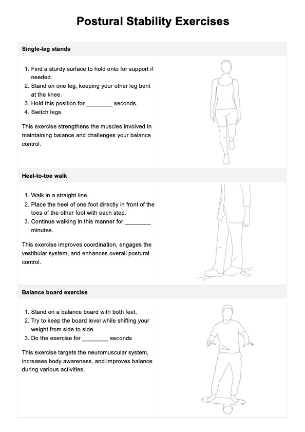
Initial treatment typically includes rest, ice application, and nonsteroidal anti-inflammatory drugs (NSAIDs) to reduce inflammation and manage pain. Physical therapy exercises to strengthen and stretch the muscles around the knee can also be beneficial.
Corticosteroid injections
If conservative measures are insufficient, corticosteroid injections may be considered to reduce inflammation and provide pain relief.
Surgical intervention
In cases where conservative treatments fail, arthroscopic surgery may be necessary. This minimally invasive surgical procedure often involves removing or releasing the inflamed plica to alleviate symptoms and restore knee function.
Commonly asked questions
Common symptoms include anterior knee pain, clicking or popping sounds, and pain during activities like squatting or climbing stairs. The pain often worsens with prolonged sitting or standing.
The test involves positioning the patient's knee at 30 degrees of flexion and applying medial pressure to the patella. Pain during this maneuver suggests an inflamed medial patellar plica.
Treatment typically starts with conservative measures like rest, ice, and nonsteroidal anti-inflammatory drugs (NSAIDs). Physical therapy and corticosteroid injections may also be used. If these are ineffective, arthroscopic surgery may be necessary to remove the inflamed plica.
Related Templates

Popular Templates