The gluteal muscles, often referred to as the "glutes," consist of three major muscles located in the buttocks: the gluteus maximus, gluteus medius, and gluteus minimus. Each muscle plays a crucial role in various movements of the hip and thigh, contributing to activities such as walking, running, and climbing.
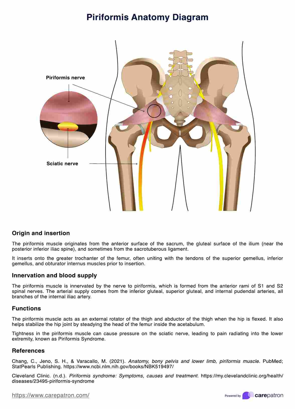
Piriformis Anatomy Diagram
What is the piriformis muscle? How does it relate to the sciatic nerve and the gluteal region? Download our free anatomy diagram to understand this crucial muscle.

Use Template
Piriformis Anatomy Diagram Template
Commonly asked questions
The piriformis muscle is a small muscle located deep within the buttocks. It extends from the anterior part of the sacrum to the upper border of the greater trochanter of the femur. It passes through the greater sciatic notch and lies beneath the gluteus maximus muscle, near the posterior margin of the pelvis.
The gluteus medius and gluteus maximus are key players in the stability and movement of the hip. The gluteus maximus is the largest and most superficial of the gluteal muscles, primarily responsible for the extension, outward rotation, and abduction of the hip. The gluteus medius, located more laterally, assists in the abduction and medial rotation of the hip and helps stabilize the pelvis during walking. The piriformis muscles work closely with these muscles to facilitate smooth and coordinated movement of the hip and thigh.


EHR and practice management software
Get started for free
*No credit card required
Free
$0/usd
Unlimited clients
Telehealth
1GB of storage
Client portal text
Automated billing and online payments