Gluteus Medius Anatomy Diagram

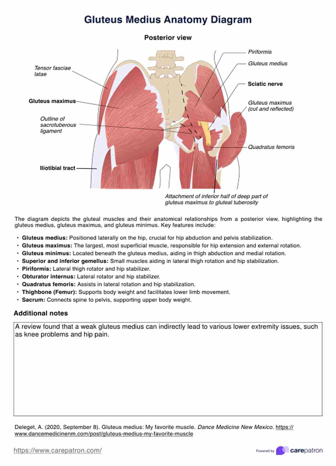
Download our free Gluteus Medius Anatomy Diagram for detailed insights into the gluteus medius, its function, and surrounding structures. Ideal for healthcare professionals.

By Olivia Sayson on Jun 24, 2024.

tick

Fact Checked by Katherine Ellison.

Use Template
Gluteus Medius Anatomy Diagram PDF Example
toolsShare uiAI icon

Introduction

The gluteus medius muscle is a crucial part of the gluteal region, playing a significant role in the movement and stability of the hip joint. It is one of the primary hip abductor muscles, essential for maintaining balance during walking, running, and other activities.

Healthcare professionals need to understand the detailed anatomy of the gluteus medius and minimus, including its relationship with surrounding structures such as the gluteus maximus, superior gluteal nerve, inferior gluteal nerve, superior gluteal artery, inferior gluteal artery, and posterior gluteal lines. This knowledge is essential for diagnosing and treating hip injuries, performing surgeries, and planning rehabilitation exercises.

Printable Gluteus Medius Anatomy Diagram PDF

Download this Gluteus Medius Anatomy Diagram to assist in identifying the source of hip pain or dysfunction.

How does our Gluteus Medius Anatomy Diagram work?

Our limb gluteus medius muscle anatomy diagram provides a detailed posterior illustration of the muscle and its anatomical context. The diagram includes:

  • Gluteus medius muscle: Highlighted to show its origin, insertion, and function.
  • Surrounding muscles: Including the gluteus maximus and gluteus minimus.

Gluteus Medius and Minimus Anatomy Diagram example (sample)

We have created a Gluteus Medius Anatomy Diagram example to help you understand how this free template works. You may view the sample here or download it as a PDF for reference.

Download our free Gluteus Medius Anatomy Diagram example here

Gluteus Medius and Minimus Anatomy Diagram example (sample)

How will our diagram benefit healthcare professionals?

Healthcare professionals, including physical therapists, orthopedic surgeons, and sports medicine specialists, can significantly benefit from our gluteus medius anatomy diagram. The gluteal muscles' diagram is particularly useful in:

  • Diagnostic assessments: Assisting in identifying the source of hip pain or dysfunction.
  • Surgical planning: Providing a visual aid for preoperative planning and intraoperative reference.
  • Rehabilitation exercises: Guiding the development of targeted rehabilitation programs for hip injuries and post-surgical recovery.
  • Educational tool: Serving as a valuable resource for teaching anatomy to medical students and residents.

Carepatron: the best healthcare OS available

Carepatron is an advanced healthcare operating system used to streamline clinical documentation, enhance patient care, and improve practice management. Our platform includes comprehensive tools for clinical documentation, ensuring that healthcare professionals have access to accurate and organized electronic patient records.

Sign up to Carepatron and experience the best healthcare OS today!

Practice Management Software
What are the primary functions of the gluteus medius and minimus muscles?

Commonly asked questions

What are the primary functions of the gluteus medius and minimus muscles?

The gluteus medius and minimus muscles are crucial hip abductor muscles. They significantly stabilize the pelvis and lower limb during walking, running, and single-leg activities.

How does the superior gluteal nerve interact with the gluteus medius muscle?

The superior gluteal nerve innervates the gluteus medius muscle and the gluteus minimus and tensor fasciae latae muscles. This nerve arises from the posterior divisions of the L4, L5, and S1 nerve roots.

Why is it important to understand the anatomy of the gluteal region, especially for hip joint surgeries?

Understanding the anatomy of the gluteal region is vital for hip joint surgeries because it helps surgeons avoid critical structures such as the superior and inferior gluteal nerves, the superior gluteal artery, and the gluteus maximus and minimus muscles.

Join 10,000+ teams using Carepatron to be more productive

One app for all your healthcare work