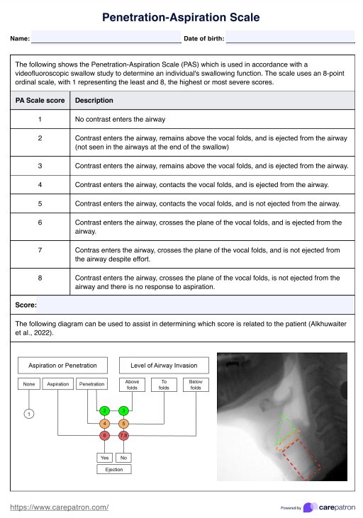
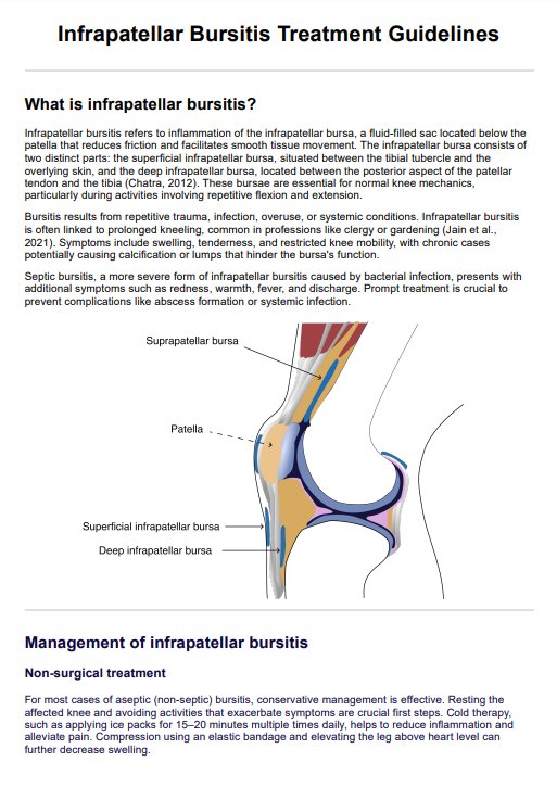
Cervical Spine Anatomy Diagram
Learn about the anatomy of the cervical spine with our free downloadable diagram. Ideal for healthcare practitioners treating neck conditions.

Fact Checked by Nate Lacson.


What is the cervical spine?
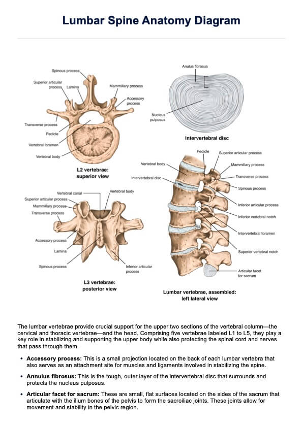
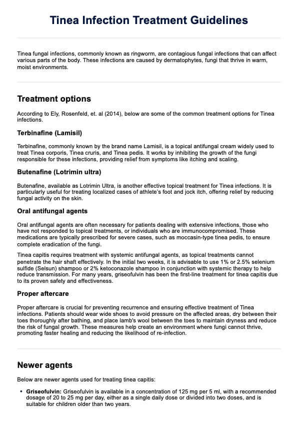
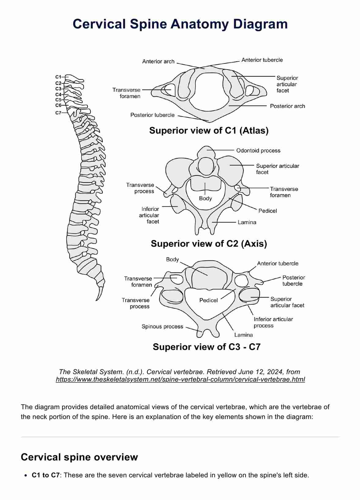
The cervical spine is the uppermost portion of the vertebral column, consisting of seven cervical vertebrae (C1-C7). Its main functions are to support the weight of the head, enable head and neck movement, and protect the spinal cord and vertebral arteries that pass through it.
The first two cervical vertebrae, the atlas (C1) and axis (C2), are specialized to allow for extensive rotation of the head. The remaining vertebrae (C3-C7) follow a more typical structure, with vertebral bodies separated by intervertebral discs that act as shock absorbers. Spinal nerves exit between each vertebral level.
The cervical spine also includes critical structures such as the anterior longitudinal ligament and posterior longitudinal ligament, which provide stability. The vertebral foramen is the passageway for the spinal cord, and injuries to this area can lead to significant spinal cord injury and chronic neck pain. Understanding the anatomy of the cervical spine is crucial for procedures like cervical spine surgery and for diagnosing conditions related to the cervical vertebrae and cervical spine ligaments.
Cervical Spine Anatomy Diagram Template
Cervical Spine Anatomy Diagram Example
What is the importance of knowing cervical spine anatomy?
A thorough understanding of cervical spine anatomy is crucial for healthcare professionals who treat conditions affecting the neck region. This knowledge is essential for:
- Diagnosing cervical spine disorders based on the location of pain and neurological symptoms
- Planning safe and effective manual therapy techniques and exercises
- Identifying contraindications and red flags prior to treatment
- Communicating findings and rationale to patients and other providers
- Recognizing potentially serious pathologies that require immediate referral
How does our Cervical Spine Anatomy Diagram work?
Here are the steps on how you can effectively utilize the diagram:
Step 1: Download the template
Access the provided link to download the free PDF of the Cervical Vertebrae Anatomy Diagram. Save it to your device for easy reference.
Step 2: Review the diagram
Open the PDF and familiarize yourself with the detailed anatomical views of the cervical vertebrae, particularly C1 (Atlas), C2 (Axis), and C3-C7. Note the key features such as the anterior and posterior arches, transverse foramen, superior and inferior articular facets, and spinous processes.
Step 3: Utilize for education and diagnosis
Use the diagram to educate students, patients, or colleagues about the anatomy of the cervical vertebrae. Highlight the unique aspects of each vertebra, such as the odontoid process of C2 or the transverse foramina's importance for vertebral arteries. Apply the visual aid in clinical settings to help diagnose conditions related to the cervical spine, referencing specific structures and their functions.
Step 4: Incorporate into reports and presentations
Include the diagram in your medical reports, presentations, or educational materials. It provides a clear, detailed visual reference to enhance understanding and communication about cervical vertebrae anatomy.
How will our diagram benefit healthcare professionals?
Here are four benefits of using our Cervical Spine Anatomy Diagram:
- Enhanced anatomical knowledge for practitioners: Our Cervical Spine Anatomy Diagram serves as an excellent reference tool, helping physical therapists, occupational therapists, chiropractors, osteopaths, orthopedic surgeons, neurosurgeons, massage therapists, athletic trainers, radiologists, and sonographers refresh their anatomical knowledge prior to patient assessment or treatment.
- Improved patient education: The diagram is a valuable resource for educating patients about their cervical spine condition. Practitioners can use it to visually explain the anatomy, the location of issues, and the rationale behind recommended interventions.
- Facilitated collaboration on complex cases: Our diagram supports effective collaboration among colleagues, especially in complex cervical spine cases. Providing a clear visual reference helps practitioners from different specialties communicate more effectively about specific anatomical structures and potential treatment approaches.
- Enhanced training for students and new graduates: The Cervical Spine Anatomy Diagram is an indispensable tool for training students and new graduates. It aids in teaching the detailed anatomy of the cervical spine, helping them to visualize and understand the relationships between different vertebrae and associated structures.
Commonly asked questions
The atlas (C1) is the first cervical vertebra. It supports the skull, allows nodding motions, and, along with the axis (C2), enables head rotation. It is crucial for the stability and mobility of the upper cervical spine.
Cervical spine ligaments, including the anterior and posterior longitudinal ligaments, ligamentum flavum, and alar and transverse ligaments, maintain stability and alignment of the cervical vertebrae. They prevent excessive movements and protect the spinal cord and spinal nerves.
- Cervical spondylosis: Degenerative changes causing neck pain and stiffness.
- Herniated disc: Disc protrusion compressing spinal nerves or cord.
- Cervical radiculopathy: Nerve root compression causing pain and numbness.
- Cervical myelopathy: Spinal cord compression causing neurological deficits.
- Whiplash: Ligament and muscle injury from a sudden force.
- Cervical spine fractures: Trauma-induced vertebrae breaks.
- Osteoarthritis: Joint cartilage degeneration causing pain and reduced mobility.
Related Templates

Popular Templates