Pisiform Fracture Test

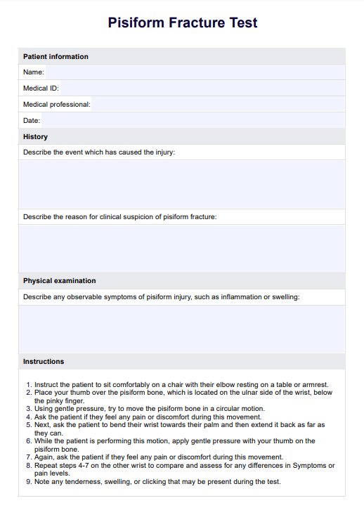
Access an easy-to-use Pisiform Fracture Test template for accurate diagnosis of pisiform injuries.

By Chloe Smith on Feb 16, 2025.

tick

Fact Checked by Ericka Pingol.

Use Template
AI IconToolbarShare ui

What are pisiform fractures?

The pisiform bone is a small, pea-shaped carpal bone located at the base of the palm on the ulnar (pinky finger) side of the wrist. It plays a crucial role in wrist stability and movement, serving as an attachment point for the abductor digiti minimi muscle and the flexor ulnaris (FCU) tendon. As one of the eight carpal bones, the pisiform is also involved in protecting the ulnar nerve along its palmar surface.

Injuries to the pisiform, such as parasagittal comminuted fractures or conditions resulting from pisiform triquetral impaction patterns, can lead to complications including ulnar nerve palsy. Severe cases may require surgical excision to alleviate symptoms and restore function.

Pisiform fractures may be caused by direct trauma or repetitive stress on the wrist, such as in sports activities like baseball, gymnastics, and rock climbing. These injuries are both rare and have symptoms that overlap with other carpal fractures, as well as being difficult to detect using standard X-rays. Consequently, diagnosis of pisiform fractures can be difficult.

Symptoms of an acute injury to the pisiform may include:

  • Swelling and tenderness on the ulnar side of the wrist
  • Difficulty or pain with gripping and grasping objects
  • Weakness in the pinky finger muscles
  • Numbness or tingling in the ring and little fingers (signs of ulnar nerve palsy)
  • Chronic wrist pain, especially with movements that involve ulnar deviation (bending the wrist towards the pinky side).

Pisiform fractures are most commonly seen in young athletes due to the repetitive stress on the wrist during sports activities. Other risk factors for pisiform fractures include:

  • Falls onto an outstretched hand, particularly with an impact on the ulnar side of the wrist
  • Direct blow or trauma to the pisiform bone or surrounding area
  • Osteoporosis (weak and brittle bones)
  • Arthritis or other degenerative joint conditions

What is a Pisiform Fracture Test?

Although not widely standardized for diagnosing pisiform fractures, the Pisiform Fracture Test involves palpating the medial aspect of the wrist where the pisiform bone is situated. Since pisiform fractures are relatively rare, it can help determine whether specialized radiographs to detect them are warranted (e.g. supinated reverse oblique wrist radiograph).

To perform the test, the examiner applies gentle pressure on the pisiform bone located near the pinky finger, moving it in a circular motion. Then, the patient is asked to bend and extend their wrist while assessing for any pain. The test is repeated on the other wrist for comparison, noting any tenderness, swelling, or clicking sounds.

Apart from manual palpation, healthcare providers should utilize various imaging techniques, such as X-rays, computerized tomography (CT) scans, or magnetic resonance imaging (MRI) to precisely confirm the diagnosis of a pisiform fracture. These imaging modalities play a crucial role in determining the fracture's exact location, type, and severity while also assessing any accompanying injuries or complications that may impact the treatment plan.

How to use our Pisiform Fracture Test template

Carepatron's simple and easy-to-use Pisiform Fracture Test template is designed to streamline the process of correctly performing and recording the Pisiform Fracture Test. Here's how it works:

Step 1: Download the template

Click "Use template" on this page to open the template in the Carepatron app, where it can be customized. Alternatively, access a ready-to-go PDF by clicking "download."

Step 2: Input patient details

Record crucial patient details at the top of the form. Record relevant medical history details, including their symptoms and the event which caused the injury. If relevant, include the reason for the assessment, e.g. if plain radiographs remain nondiagnostic.

Step 4: Perform the Pisiform Fracture Test

Follow the instructions outlined in the template to examine the pisiform bone thoroughly. During palpation, note any tenderness, swelling, or clicking sounds, as well as the location.

Step 5: Record results and findings

Record your observations and results from the examination in the designated sections of the template. This will serve as a clear record of your assessment for future reference.

Step 6: Conduct imaging or further tests

Conduct imaging or further tests to confirm the diagnosis and extent of the fracture. You can also use the template to record and include these results in the patient's file.

Benefits of using the Pisiform Fracture Test template

This template has been specifically formulated to aid healthcare professionals in testing for fractures of the pisiform. Here's some of the key advantages:

Streamlined diagnosis of pisiform fractures

This template has been designed to streamline the diagnostic process of assessing for pisiform fractures. With easy-to-follow formatting and instructions, this template efficiently guides healthcare professionals through collecting relevant information and performing the test.

Increased accuracy

This template has comprehensive step-by-step instructions to ensure the test is performed and interpreted accurately. This helps to increase diagnostic accuracy, ensuring patients receive appropriate and effective treatment.

Accurate documentation

This Pisiform Fracture Test template has been designed to guide healthcare professionals to record all key details of a patient's diagnostic assessment. Thorough record-keeping facilitates shared understanding of a patient's condition between healthcare team members and the patient themselves, as well as holding professionals accountable for providing appropriate standards of medical care.

Pisiform fracture treatments

Treatment options for pisiform fractures may vary depending on the type and severity of the fracture, as well as any accompanying injuries or conditions. Some common treatment methods include:

  • Immobilization: For non-displaced or minimally displaced fractures, a splint, cast, or brace may be used to keep the wrist in a stable position while it heals.
  • Medications: Pain relievers and anti-inflammatory medications may be prescribed to manage pain and reduce inflammation.
  • Physical therapy: Rehabilitation exercises and physical therapy may be recommended to strengthen the wrist and restore range of motion after the fracture has healed.
  • Excision of the pisiform: Pisiform excision surgery may be necessary in cases of severe or unstable fractures, or if non-surgical treatments have been unsuccessful.
    • Open reduction and internal fixation (ORIF) surgery may be necessary for a displaced pisiform fracture.
    • Surgery to decompress the ulnar nerve may be necessary if fracture fragments have caused ulnar nerve damage. Pisiform-fracture-associated ulnar nerve palsy may take around 8-12 weeks following surgery to resolve.
What is a pisiform fracture?

Commonly asked questions

What is a pisiform fracture?

Isolated pisiform fractures are rare injuries in which a direct blow or trauma to the wrist causes a break or crack in the small, pea-shaped bone located on the ulnar side of the wrist.

How do pisiform fractures typically occur?

Pisiform fractures are often caused by a direct blow or impact to the palm of the hand, such as during a fall onto an outstretched hand. An acute transverse fracture of the pisiform often occurs alongside other injuries to the carpal bones.

What are the common symptoms of a pisiform fracture?

Symptoms may include ulnar sided wrist pain and tenderness, difficulty moving the wrist, and swelling or bruising around the area. Some patients may also experience numbness or tingling in the 4th and 5th fingers, especially if there is concomitant injury to the ulnar nerve. Symptoms often overlap with other types of carpal fractures.

How are pisiform fractures diagnosed?

Diagnosing a pisiform fracture involves a comprehensive physical examination, including a review of medical history and the use of imaging techniques. Standard X-rays, such as an oblique radiograph with the wrist supinated at 45 degrees, are commonly employed; however, plain X-rays may not always detect pisiform fractures. In such cases, advanced imaging like CT scans or MRIs can help identify fractures, including parasagittal comminuted patterns involving the pisiform.

Join 10,000+ teams using Carepatron to be more productive

One app for all your healthcare work