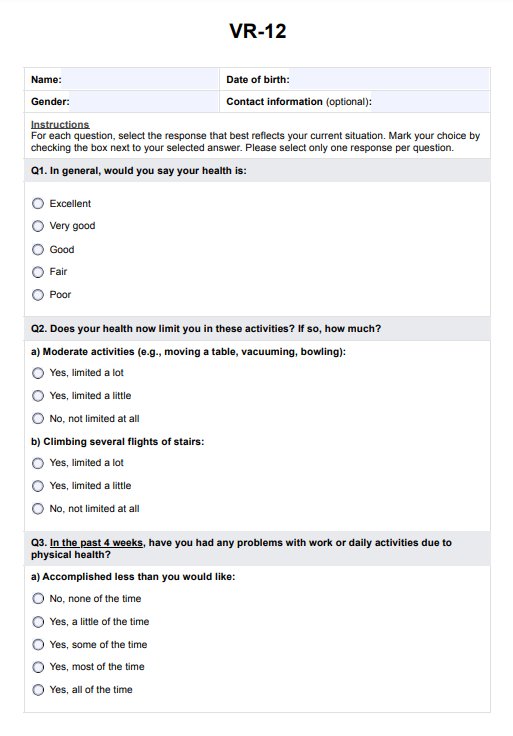
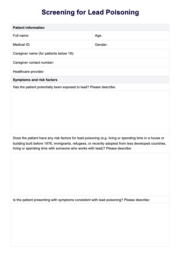
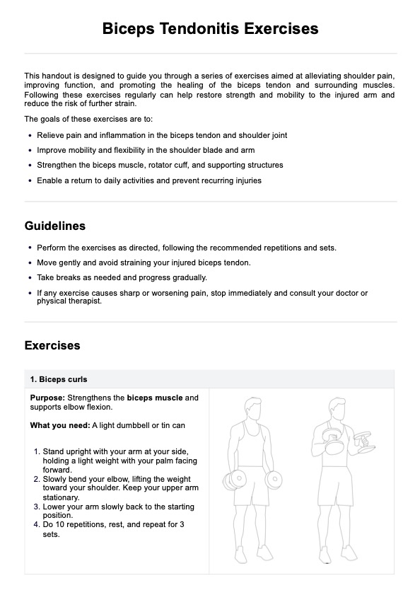
Wrist Radiograph for Carpal Bone Fractures Worksheet
Explore our detailed Wrist Radiograph for Carpal Bone Fractures Worksheet for insights into effectively diagnosing and treating wrist fractures.

Fact Checked by Ericka Pingol.


What are the different views on a wrist radiograph?
Wrist radiographs are commonly used in medical imaging to assess injuries and conditions affecting the wrist joint and surrounding areas. These X-rays provide critical information about bone alignment, fractures, and signs of degenerative diseases. Several standard views are taken in wrist radiography, each offering a unique perspective and useful for diagnosing different issues.
Posteroanterior (PA) view
The posteroanterior view, often abbreviated as PA, is the standard and initial view taken during wrist radiography. In this view, the X-ray beam passes from the back (posterior) of the hand to the front (anterior). The patient places their arm and hand palm-down on the X-ray table, ensuring the wrist is in a neutral position. This view is essential for evaluating the overall bone structure, including the distal radius, ulna, and carpal bones. It is particularly useful for identifying fractures in the radius and ulna and assessing bone density and joint spaces.
Lateral view
The lateral view is taken with the X-ray beam passing from one side of the wrist to the other, usually from the thumb side (radial) to the little finger side (ulnar). The patient’s hand is positioned so that the thumb is pointing upwards. This view provides a side profile of the wrist and is crucial for assessing the alignment of the radius and ulna with the carpal bones. It helps evaluate the palmar or dorsal displacement of acute wrist trauma or fractures and the integrity of the dorsal and palmar rims of the wrist.
Oblique view
The oblique view is achieved by rotating the wrist at an angle, usually about 45 degrees, while the X-ray is taken. Depending on the direction of the wrist rotation, this view can be medial or lateral. It provides a diagonal perspective of the wrist bones and is particularly helpful for visualizing the scaphoid bone, one of the carpal bones that is frequently fractured. The oblique view can reveal fractures or abnormalities, such as occult scaphoid fractures that are not visible in the PA or lateral views.
Special views
In addition to the standard views, there are several specialized views used to highlight specific areas of the hand and wrist:
- Scaphoid view: Specifically designed to focus on the scaphoid bone, this view requires the wrist to be ulnar deviated. It is used when a scaphoid fracture is suspected but not clearly visible in other views.
- Carpal tunnel view: This view visualizes the bony and soft tissue structures of the carpal tunnel. The hand is positioned to look directly into the tunnel, which helps assess conditions like carpal tunnel syndrome.
- Ulnocarpal stress view: This view is performed while applying stress or pressure to the ulnar side of the wrist. It helps assess the stability of the ulnar aspect of the wrist and detect subtle shifts that may indicate ligament damage.
Each of these views provides vital information for a comprehensive assessment of the wrist, aiding in accurate diagnosis and effective treatment planning.
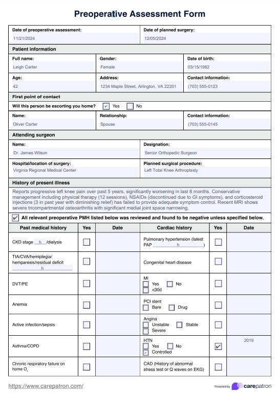
Wrist Radiograph for Carpal Bone Fractures Worksheet Template
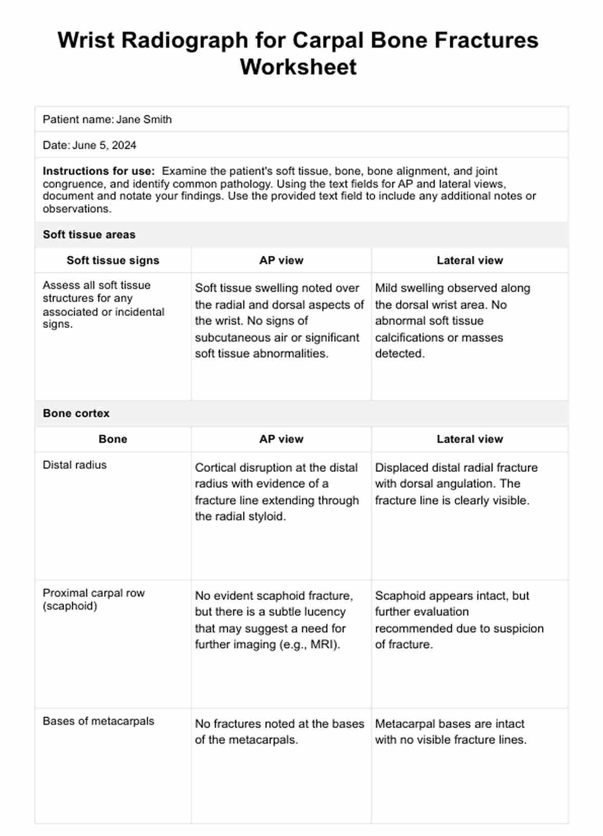
Wrist Radiograph for Carpal Bone Fractures Worksheet Example
What does an X-ray of the wrist show?
An X-ray of the wrist provides a detailed image of the bones that make up the wrist joint, including the distal ends of the radius, the distal ulna (the two long forearm bones), and the eight small carpal bones that form the wrist itself. X-rays effectively detect various abnormalities such as fractures, signs of osteoarthritis, and changes in bone density.
They can also help assess the alignment of the wrist bones and the joint spacing, which is crucial for diagnosing conditions like rheumatoid arthritis or other degenerative diseases. Furthermore, wrist X-rays are used to evaluate any suspected abnormalities in the soft tissues and ligaments surrounding the wrist, although soft tissues and ligament injury are not as clearly visible as bones on an X-ray.
Can you see a wrist fracture on an X-ray?
Yes, wrist fractures are commonly visible on an X-ray, the primary diagnostic tool for their detection. X-rays of wrist pain can clearly show breaks in the continuity of the bone, ranging from simple fractures that may require minimal treatment to complex ones that might need surgical intervention.
The visibility of a fracture depends on its location and orientation; for example, some scaphoid fractures might not be immediately apparent and could require special views or follow-up X-rays. Overall, an X-ray provides crucial information on the type and extent of the fracture, aiding in appropriate treatment planning.
What is a distal radius fracture?
A distal radius fracture is one of the most common types of wrist fractures and occurs near the wrist joint at the distal end of the radius, the larger of the two forearm bones. This type of distal pole fracture typically happens when someone falls onto an outstretched hand, which can cause the bone to break on the lower end distal radius fractures just above where it connects to the wrist.
Symptoms include immediate pain, tenderness, swelling, and sometimes a visible wrist deformity, suggesting a displaced bone. Treatment varies based on the severity of the fracture, ranging from casting and immobilization for simple fractures to surgical intervention for more complex or displaced fractures.
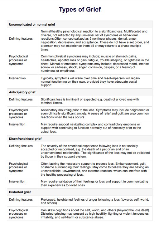
What is the proximal carpal row?
The proximal carpal row refers to the first row of carpal bones closest to the forearm, within the two rows of bones that constitute the wrist. It includes the following four other carpal bones: the scaphoid, lunate, triquetrum, and pisiform.
These bones are fundamental in the complex movements of the wrist, allowing joint space for the flexion, extension, and rotational movements of the hand. Each bone in the proximal row articulates with the distal ends of the radius and ulna and plays a crucial role in transmitting the forces from the hand to the forearm.
Are fractures on the carpal bones deadly?
Fractures of the carpal bones are generally not deadly, but they can lead to complications if not treated properly. The most common fractured carpal bone is the scaphoid, which can sometimes lead to nonunion or avascular necrosis (death of bone tissue due to a lack of blood supply) if not diagnosed and managed correctly.
These complications can result in chronic pain, reduced range of motion, and long-term disability. However, with timely and appropriate treatment, most people recover well from carpal bone fractures without life-threatening consequences.
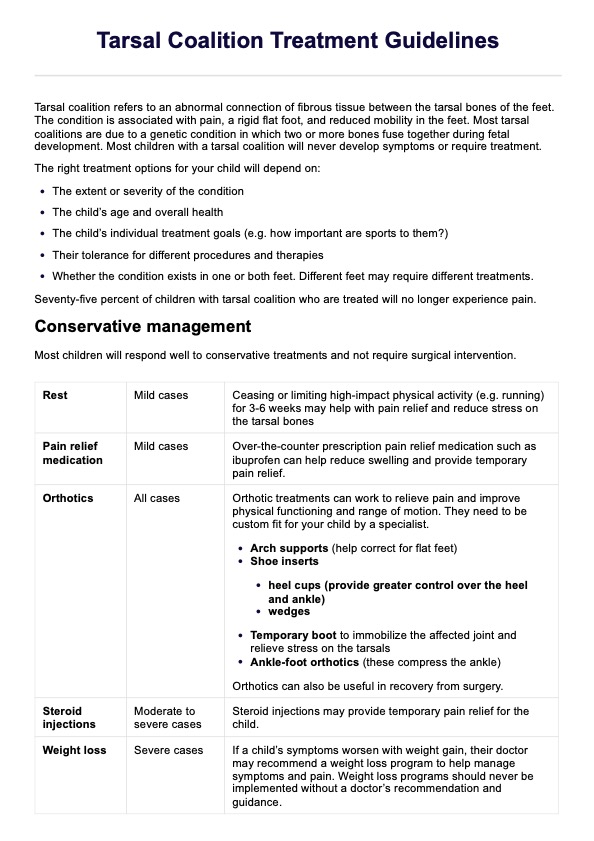
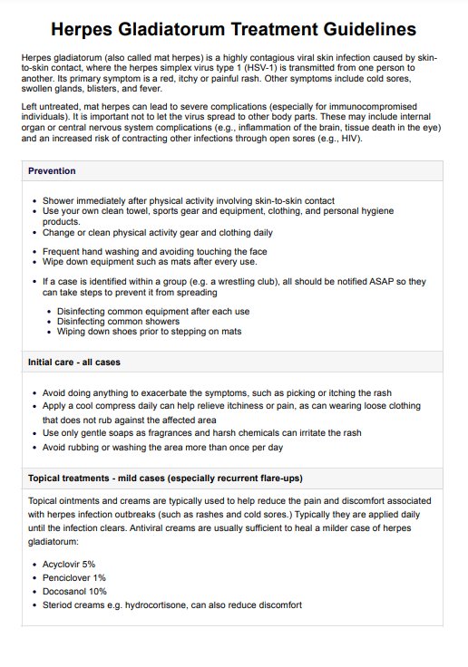
How to use our Radiograph for Carpal Bone Fractures Worksheet
This worksheet is designed to guide healthcare professionals through the process of assessing soft tissue areas, bone cortex, bony alignment, joint congruency, and identifying common pathologies. By following these steps meticulously, one can ensure a comprehensive evaluation, leading to accurate diagnosis and appropriate management of carpal bone fractures:
Step 1: Assess soft tissue areas
Examine the soft tissue areas in both the AP and lateral views. Assess all soft tissue structures for any associated or incidental signs. Specifically, look for indications of swelling, effusion, or soft tissue injuries that may accompany bone fractures or dislocations. This initial step is crucial for identifying any underlying issues that might not be immediately apparent in the bone structures.
Step 2: Examine the bone cortex
Next, focus on the bone cortex, starting with the distal radius. In the AP view, check the distal radial articular surface to ensure proper cupping of the carpals and verify that the radial inclination angle is between 15-25 degrees. The radial surface should appear smooth. In the lateral view, confirm that the palmar tilt of the articular surface is within 10-25 degrees, ensuring the volar tilt is normal.
Move on to the proximal carpal row, specifically the scaphoid bone. In the AP view, look for any signs of fracture or dislocation. Verify that the scaphoid bone aligns properly with the distal radius in the lateral view. Lastly, examine the bases of the metacarpals. In the AP view, check for fractures or dislocations, and in the lateral view, ensure the metacarpals align correctly with the carpals.
Step 3: Evaluate bony alignment
Proceed to evaluate the bony alignment, starting with the carpal arcs in the AP view. Check the articular surfaces of both the proximal and distal carpal rows for three smooth arcs and ensure the spacing between all carpal bones is 1-2 mm. Then, in the lateral view, verify the carpal alignment by checking that the distal radius, lunate, and capitate align in a straight line. Look for any signs of lunate or perilunate dislocation.
Step 4: Check joint congruency
Examine the joint congruency, beginning with the carpometacarpal articulation. In the AP view, check that the joint space between the carpals and metacarpals is 1-2 mm. In the lateral view, verify that the joint space remains within normal limits, ensuring proper joint congruency and alignment.
Step 5: Identify common pathology
Look for common pathologies, starting with fractures. In the AP view, check for dorsal angulation in Colles fractures and confirm whether the fracture is intra-articular or extra-articular. For Smith fractures, check for palmar angulation in the lateral view and verify that the fracture is extra-articular. Examine the scaphoid for signs of fracture, particularly through its waist, and note if the fracture is radiographically occult.
For triquetral fractures, look for dorsal chip fractures and verify if these fractures are radiographically occult. Finally, inspect other carpal bones (lunate, capitate, hamate, trapezium, trapezoid, and pisiform) for signs of fractures and confirm if they are radiographically occult if suspected.
Commonly asked questions
A wrist radiograph helps identify breaks or abnormalities in the carpal bones, which is crucial for diagnosing fractures and planning appropriate treatment strategies.
The scaphoid bone is the most frequently used fracture displacement of a fractured carpal bone, and it is often clearly visible on a wrist X-ray, especially when specific scaphoid views are used.
Common signs of a carpal bone fracture on an X-ray include visible cracks or breaks in the bone, changes in bone alignment, and abnormal spacing between the carpal bones.
Related Templates

Popular Templates


-template.jpg)