Electromyography
Discover the power of Electromyography (EMG) tests! Explore their many uses and learn how to interpret results with confidence.


What is an Electromyography Test?
Electromyography, often abbreviated as EMG, is a diagnostic procedure used in neurology and sports medicine to assess the health and function of muscles and the nerves that control them. This test plays a crucial role in identifying and diagnosing various neuromuscular disorders. It is commonly employed to aid in evaluating conditions such as muscular dystrophy, carpal tunnel syndrome, amyotrophic lateral sclerosis (ALS), and more.
The EMG test involves using a specialized instrument known as an electromyograph, which records electrical activity in muscles. During the procedure, fine needle electrodes are inserted into the targeted muscle groups under examination. These electrodes detect and record the electrical signals generated by muscle contractions and the nerves that control them. The patient is usually asked to perform specific movements or contract their muscles to elicit a response, allowing the healthcare provider to analyze the data.
The primary objectives of an EMG test include assessing muscle function, identifying nerve damage, and differentiating between nerve and muscle disorders. The results of an EMG test provide valuable information to physicians, helping them formulate treatment plans and monitor the progression of neuromuscular diseases. While the procedure may cause mild discomfort due to the insertion of electrodes, it is generally safe and well-tolerated.
EMG is an essential tool in assessing neuromuscular function and is crucial in improving the diagnosis and management of a wide range of neurological and musculoskeletal conditions.
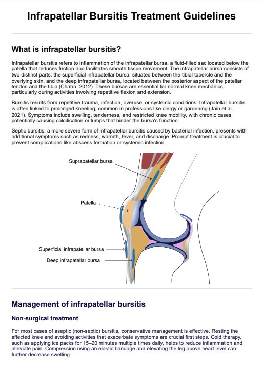
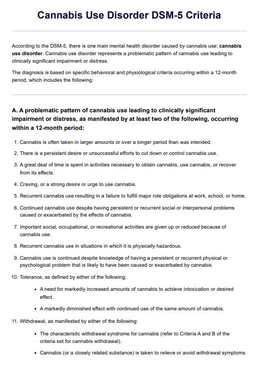
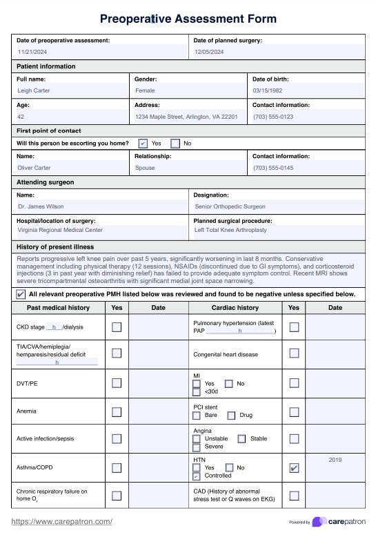
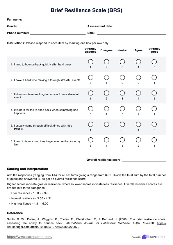
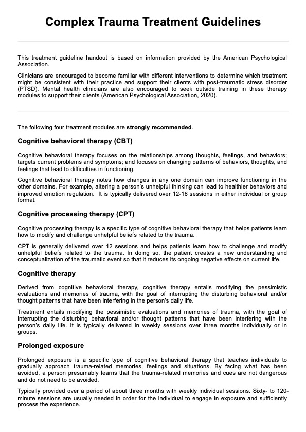
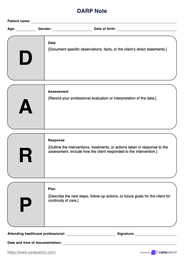
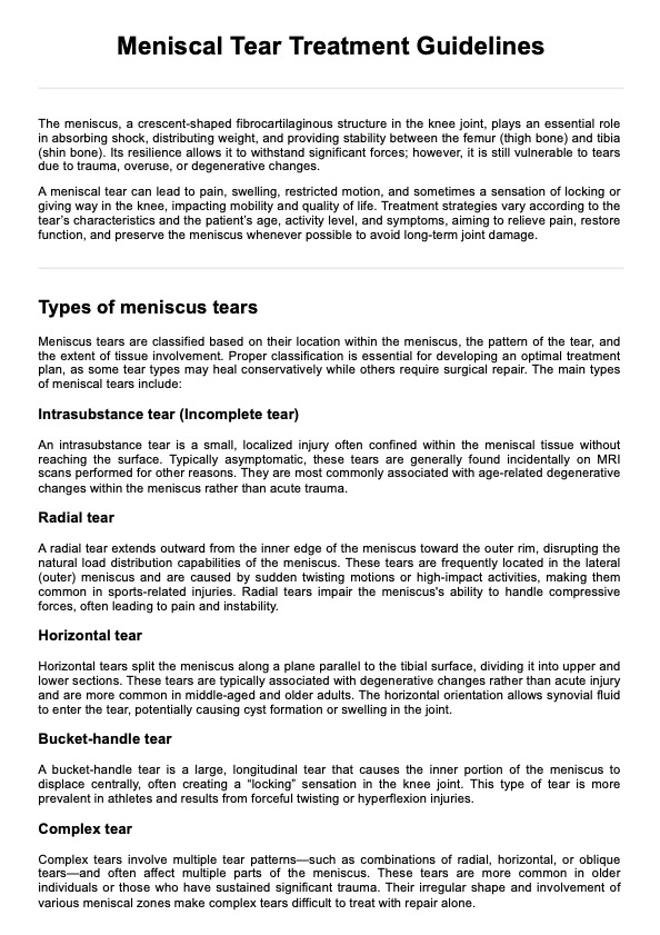
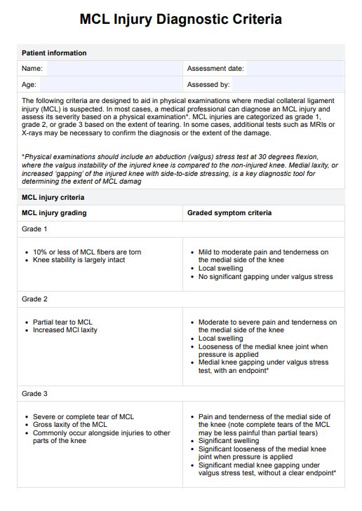
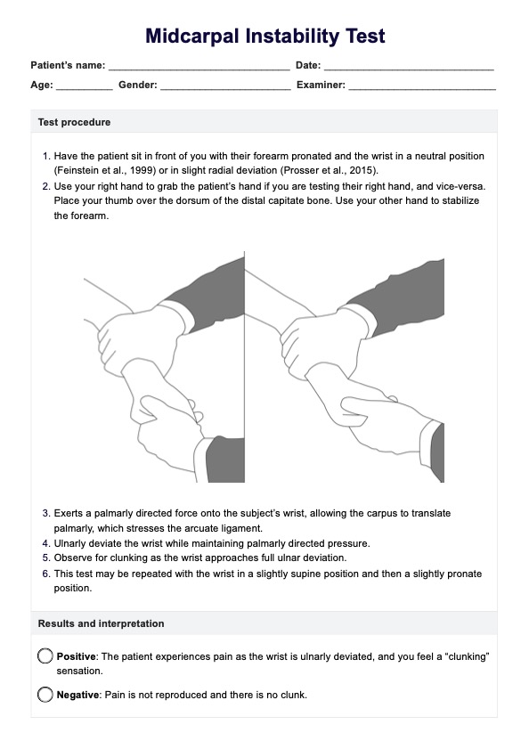
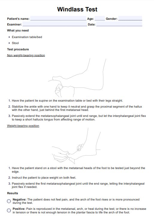
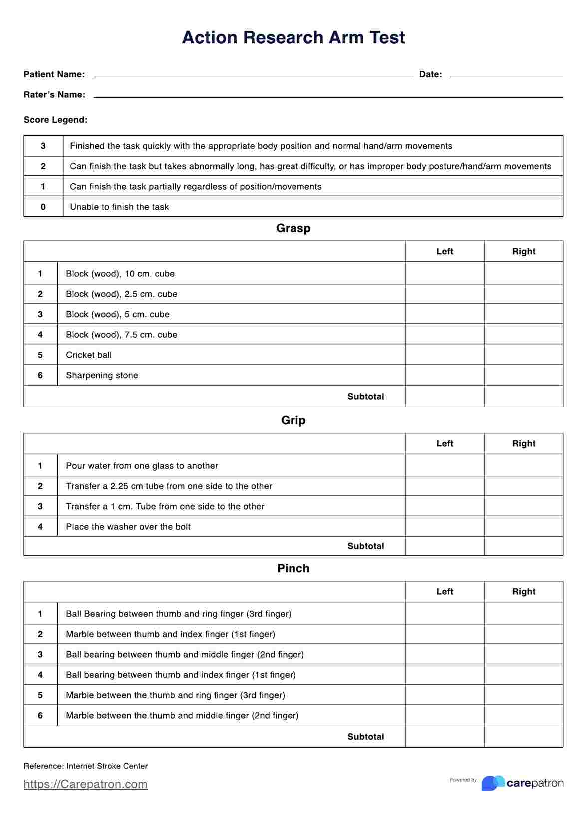
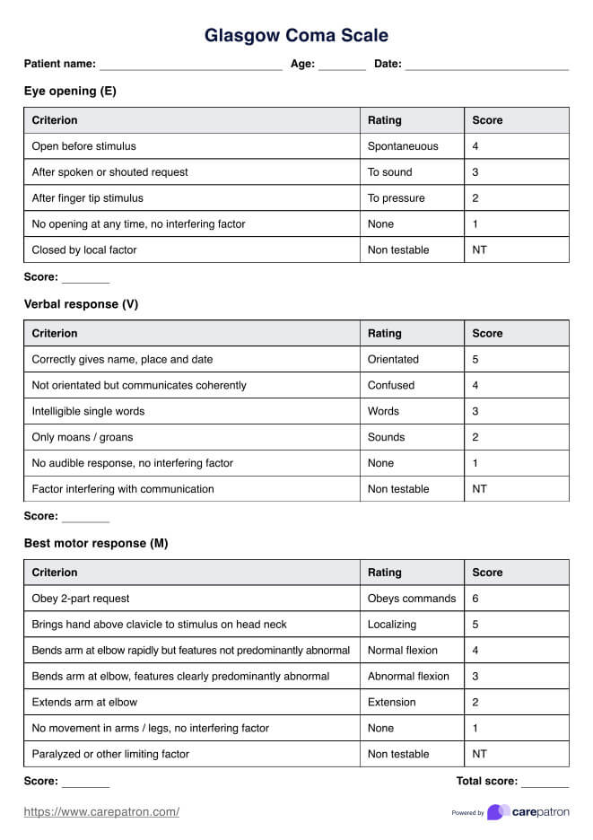
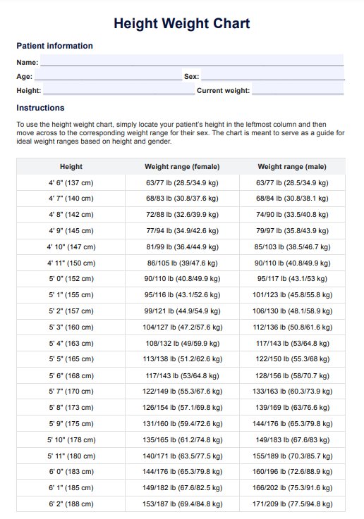
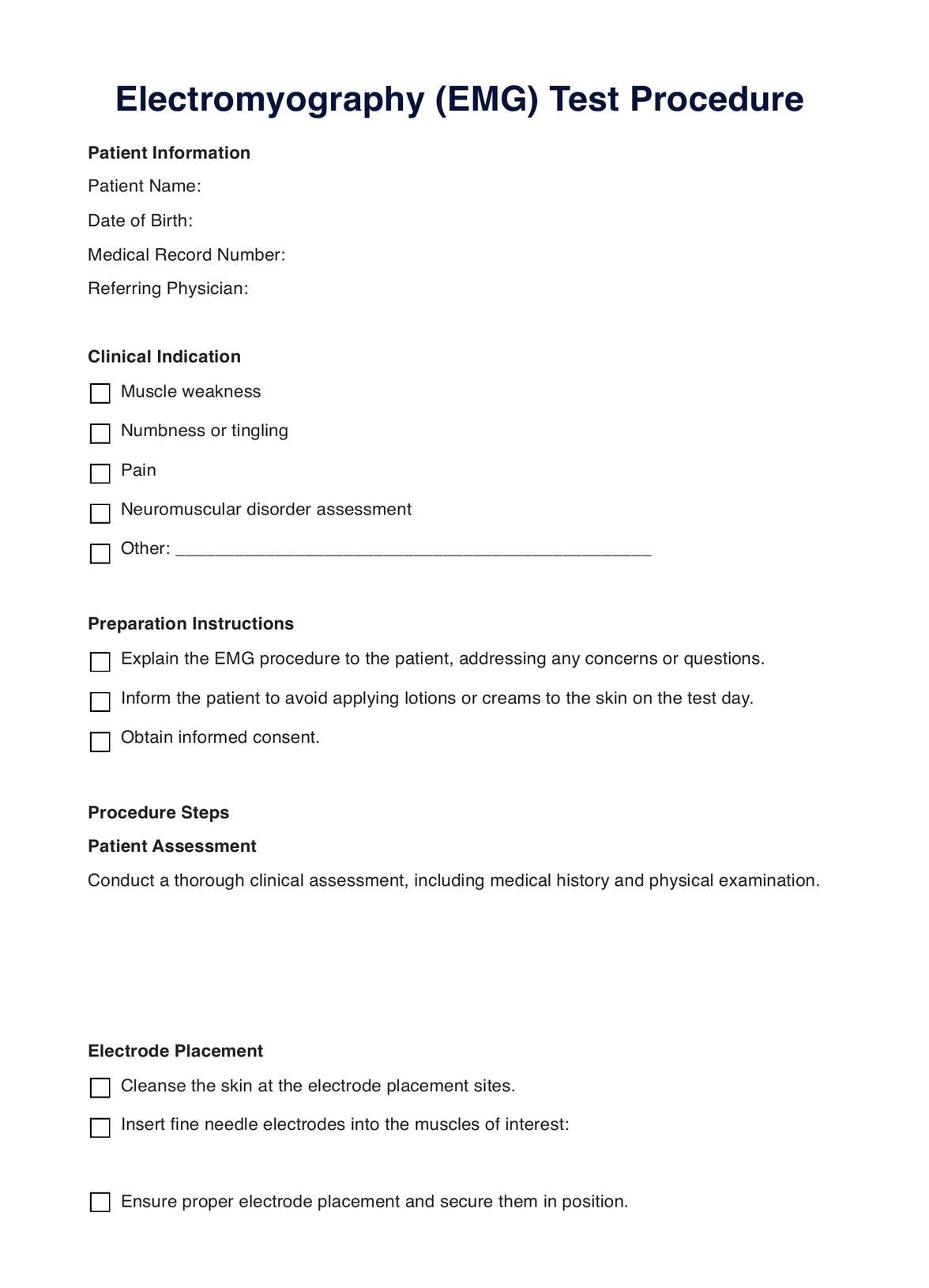
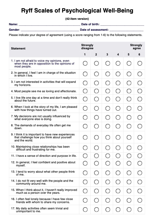
Electromyography Template
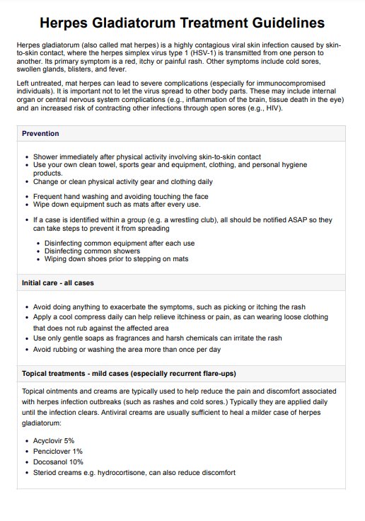
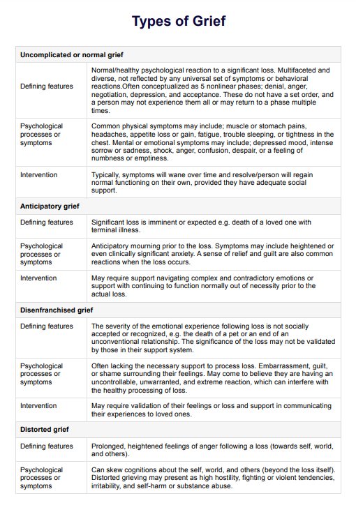
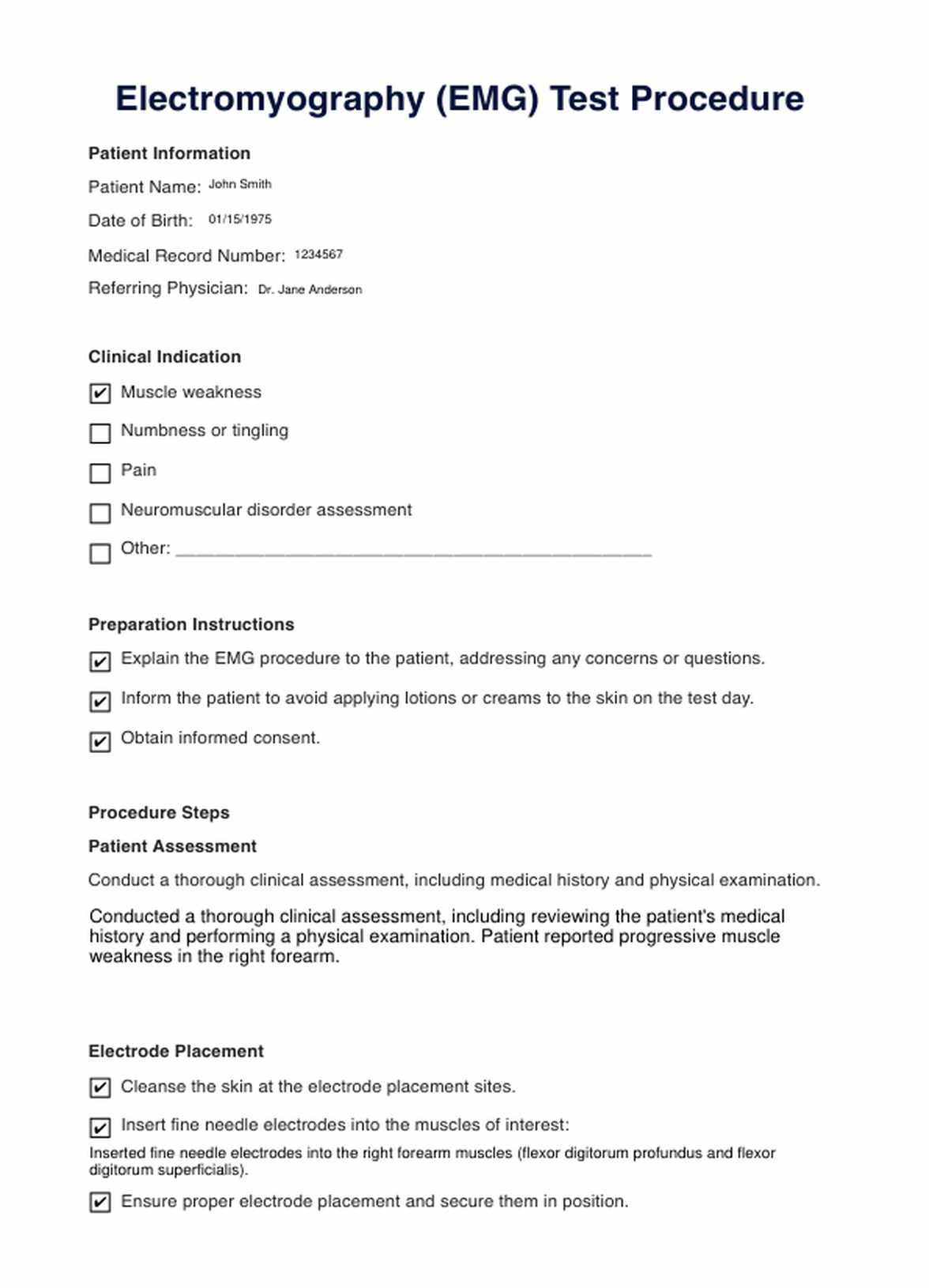
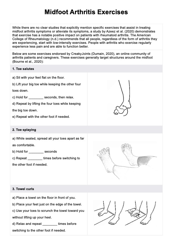
Electromyography Example
How Does it Work?
1. Patient Preparation
The patient is typically briefed on the procedure, its purpose, and what to expect. They may be asked to avoid applying lotions or creams to the skin on the day of the test, as these can interfere with electrode placement.
2. Electrode Placement
A trained healthcare provider, often a neurologist or a specially trained technician, positions fine needle electrodes into specific muscles of interest. These electrodes act as sensors to detect electrical activity in the muscles.
3. Muscle Activity Assessment
Depending on the specific muscles being tested, the patient is instructed to perform various muscle contractions or movements. As the patient contracts their muscles, the EMG records the electrical signals generated by these contractions.
4. Nerve Conduction Study (Optional)
Sometimes, a nerve conduction study (NCS) may be performed alongside EMG. NCS involves delivering small electrical pulses to stimulate nerves and assess how well they transmit signals to the muscles.
5. Data Collection
The electromyograph records the electrical activity through waveforms or tracings. These data provide information about the amplitude, frequency, and duration of muscle contractions and the integrity of nerve-muscle connections.
6. Interpretation
The recorded data are analyzed by a trained specialist who interprets the findings. Abnormalities in muscle activity or nerve function are identified and used to make a diagnosis.
7. Report and Diagnosis
The results of the EMG are documented in a report that includes the diagnosis or assessment of neuromuscular function. This report is shared with the referring physician to guide treatment decisions.
8. Follow-up and Treatment
Based on the EMG findings, the patient's healthcare provider develops a treatment plan, including physical therapy, medications, or further testing, depending on the diagnosis.
When Would you use this Test?
The Electromyography (EMG) Test is a valuable diagnostic tool with many applications across various medical specialties. Healthcare practitioners, including neurologists, orthopedic surgeons, physiatrists, and sports medicine specialists, rely on the EMG test to assess neuromuscular function and diagnose various conditions. Here are some key scenarios in which this test is appropriate:
- Neuromuscular Disorders: EMG is commonly used to diagnose and monitor conditions such as muscular dystrophy, myasthenia gravis, and amyotrophic lateral sclerosis (ALS), as it helps identify muscle and nerve abnormalities.
- Nerve Compression Syndromes: Orthopedic surgeons and neurologists use EMG to diagnose conditions like carpal tunnel syndrome, cubital tunnel syndrome, and sciatica, which involve nerve compression.
- Peripheral Neuropathy: Neurologists and internists employ EMG to assess and pinpoint the cause of peripheral neuropathies, including diabetic neuropathy and autoimmune neuropathies.
- Spinal Cord Disorders: EMG is a valuable tool in diagnosing conditions affecting the spinal cord, such as radiculopathy and myelopathy.
- Muscle Disorders: Physicians use EMG to evaluate myopathy, muscle inflammation (myositis), and muscle weakness of unknown origin.
- Sports Medicine: Sports medicine specialists and physical therapists use EMG to assess muscle function and guide rehabilitation after sports-related injuries.
- Preoperative Assessment: Surgeons may request EMG testing before surgical procedures involving nerves or muscles to assess baseline function and plan the surgery effectively.
- Work-Related Injuries: Occupational medicine specialists may use EMG to evaluate and manage work-related neuromuscular injuries, helping with return-to-work decisions.
- Evaluation of Tremors and Movement Disorders: Neurologists utilize EMG to assess tremors and involuntary movements in conditions like Parkinson's disease and essential tremors.
- Unexplained Symptoms: When patients present with unexplained muscle weakness, pain, or numbness, EMG can help uncover the underlying cause.
What do the Results Mean?
Interpreting EMG test results is crucial to diagnosing neuromuscular and musculoskeletal conditions. The test provides valuable information about the electrical activity in muscles and nerves, helping healthcare professionals make accurate diagnoses and treatment decisions. Here are common EMG test results and what they typically mean:
- Normal Results: A normal EMG result indicates that the electrical activity in the tested muscles and nerves falls within expected ranges. This suggests that there is no significant neuromuscular dysfunction or abnormality.
- Denervation: Abnormal EMG findings may include signs of denervation, which indicates damage to the nerve supply to a muscle. Denervation is often seen as a spontaneous activity in the form of fibrillation potentials and positive sharp waves. It may suggest conditions like motor neuron diseases or nerve injuries.
- Myopathy: EMG can also reveal myopathic changes, which indicate muscle disease or injury. These changes are characterized by abnormal muscle activity during voluntary contraction and may indicate muscular dystrophy or inflammatory myopathies.
- Nerve Compression: In nerve compression syndromes like carpal tunnel syndrome, EMG may show slowed nerve conduction velocities and abnormal motor unit potentials, reflecting nerve irritation or damage.
- Radicular Abnormalities: EMG can identify radicular abnormalities caused by herniated discs or spinal stenosis. This is often characterized by abnormal nerve conduction studies or muscle activity patterns that suggest nerve root compression.
- Neuropathy: EMG can detect neuropathic conditions like diabetic neuropathy, where there may be reduced nerve conduction velocities and abnormal motor unit potentials, indicating nerve damage.
- Myasthenia Gravis: In myasthenia gravis, a neuromuscular junction disorder, EMG may reveal fatigable muscle responses during repetitive nerve stimulation.
EMG results require clinical correlation and further testing to determine the underlying cause. Findings guide treatment decisions. Consult a healthcare provider for a comprehensive understanding of results and management plans.
Research & Evidence
The history of electromyography dates back to the late 19th century when scientists and physicians began exploring the electrical activity of muscles and nerves. Luigi Galvani's pioneering experiments with frog legs in the late 18th century laid the foundation for understanding the relationship between electricity and muscle contraction. However, in the late 19th century, technological advancements allowed for more precise measurements of muscle electrical activity.
In 1929, the first modern EMG machine was developed by Edgar Adrian and Sir Charles Sherrington, both of whom received the Nobel Prize for their contributions to neurophysiology. This invention marked a significant milestone in the field, enabling more accurate and detailed muscle and nerve activity recordings.
Over the decades, EMG has continued evolving with advancements in electronics and computer technology. Today's EMG equipment is highly sophisticated, allowing for precise recordings and analysis of neuromuscular function.
Research and evidence supporting the use of EMG are abundant and have contributed to its widespread adoption in clinical practice. Studies have demonstrated the utility of EMG in diagnosing a wide range of neuromuscular disorders, including peripheral neuropathies, myopathies, motor neuron diseases, and nerve compression syndromes. EMG is also crucial in differentiating between nerve and muscle pathologies, aiding in selecting appropriate treatment strategies.
Furthermore, EMG research has expanded to include applications in sports medicine, physical therapy, and rehabilitation. It helps athletes and patients recover from injuries, optimize muscle function, and enhance performance.
References
- Cohen, L. (n.d.). How can EMG and NCV tests help you?: Dr. Lenny Cohen: Neurology. Dr. Lenny Cohen. https://www.chicagoneurodoc.com/blog/how-can-emg-and-ncv-tests-help-you
- Eidelson, S. G., MD. (2023, February 13). What Is Electromyography? Health Central. https://www.healthcentral.com/condition/back-pain/electromyography-emg
- Eske, J. (2020, February 19). What to know about electromyography (EMG) tests. https://www.medicalnewstoday.com/articles/emg-test
- Moores, D. (2018, March 20). Electromyography (EMG). Healthline. https://www.healthline.com/health/electromyography
- Spine, M. B. &. (n.d.). Electromyography (EMG) & Nerve conduction studies. https://mayfieldclinic.com/pe-emg.htm
- TheAANEM. (2013, September 17). What to expect during nerve conduction study and EMG test [Video]. YouTube. https://www.youtube.com/watch?v=GalU9SWiYic
Commonly asked questions
Electromyography (EMG) tests are typically requested by healthcare professionals, such as neurologists, orthopedic surgeons, and physiatrists, to assess neuromuscular and nerve function.
EMG tests are used to evaluate muscle and nerve function, diagnose neuromuscular disorders, identify nerve compression or damage, and assess a wide range of conditions causing muscle weakness, pain, or numbness.
EMG tests involve inserting fine needle electrodes into specific muscles to record electrical activity during muscle contractions. This data helps diagnose neuromuscular and musculoskeletal conditions.
The duration of an EMG test varies but typically takes between 30 minutes to an hour, depending on the number of muscles being tested and the case's complexity.









-template.jpg)
