Brain Diagrams
Looking for a brain diagram you can use as a resource? Click here for a free template and suggestions on how to use it!


What is a Brain Diagram?
All practitioners who have gone through medical and nursing school are undoubtedly familiar with the central nervous system, particularly the brain and spinal cord. However, for a refresher, the Brain Diagram is an illustration that helps practitioners learn more about the brain structure and the names and corresponding locations of the different parts of the brain.
The appearance of a Brain Diagram can vary depending on its source or purpose. Diagrams may depict the brain from a top or side view, emphasizing its external or internal parts. The level of detail can be tailored to the practitioner's specific requirements, ranging from a general overview to a more intricate representation, which might extend to the brain's connection with the peripheral nervous system.
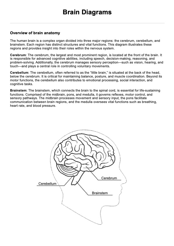
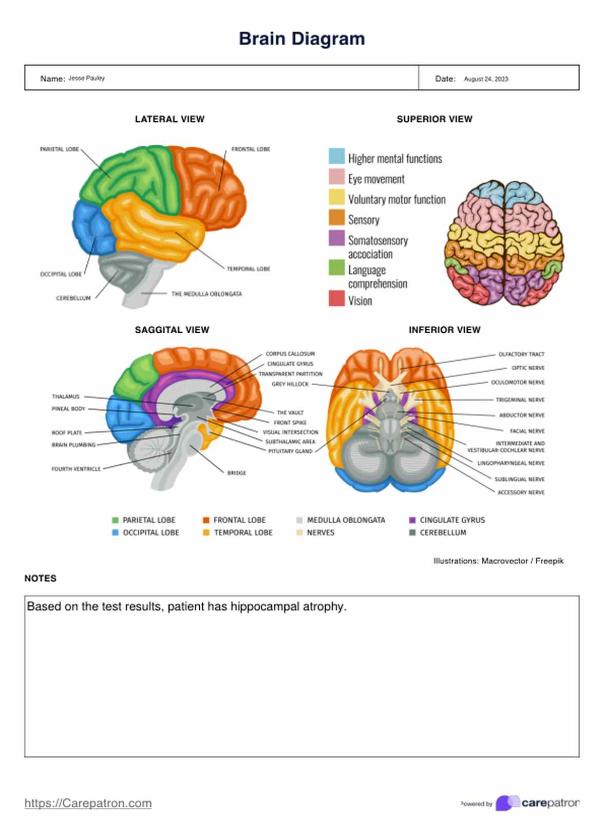
Brain Diagrams are helpful mostly to students. And though that is true, it can also be helpful to medical professionals. We prepared a Brain Diagram complete with an illustration of the brain and the corresponding labels for each part. We've also included a dedicated space for any notes you may have for yourself or your patient.
Brain Diagrams Template
Brain Diagrams Example
What is usually included in a Brain Diagram?
A comprehensive human Brain Diagram typically includes several key components that provide a detailed overview of the brain's structure and organization. Understanding these elements is crucial for healthcare practitioners when interpreting neurological findings and explaining brain function to patients.
Major brain structures
A Brain Diagram usually depicts the four main lobes of the cerebral cortex: frontal, parietal, temporal, and occipital lobes. These lobes are separated by distinct fissures and sulci, such as the central sulcus, which divides the frontal and parietal lobes. The diagram also shows the two cerebral hemispheres, separated by the corpus callosum.
Subcortical structures, including the basal ganglia, thalamus, hippocampus, and pituitary gland, are often illustrated beneath the cortex. The cerebellum, located at the base of the brain, is another prominent feature in most diagrams.
Brain stem components
The brain stem is typically depicted, showing its three main parts: the midbrain, pons, and medulla oblongata. This region is crucial as it connects the brain to the spinal cord and contains nuclei for several cranial nerves.
Functional areas
Many Brain Diagrams highlight specific functional areas within the lobes of the brain. For instance, the primary motor cortex in the frontal lobe, the primary somatosensory cortex in the parietal lobe, the primary auditory cortex in the temporal lobe, and the primary visual cortex in the occipital lobe are often labeled. The prefrontal cortex, important for executive functions, is also commonly highlighted.
Limbic system
Components of the limbic system, such as the amygdala, hippocampus, and hypothalamus, are often featured due to their roles in emotion, memory, and maintaining body temperature.
Ventricular system
The ventricular system, consisting of four interconnected cavities within the brain that produce and circulate cerebrospinal fluid, is sometimes included in more detailed diagrams.
How does it work?
Practitioners can use this Brain Diagram template by following these steps:
Step 1: Download the template
Access and download our printable Brain Diagram template by either:
- Clicking the “Use Template” or “Download Template” button above
- Searching the “Brain Diagram” in Carepatron's template library on our app or website
Step 2: Use the resource
Once you have a copy, you can use the resource in any way. For ideas on how and when to use this template, proceed to the “When would you use this Template?” section below.
Step 3: Write down notes
Since we've provided a space at the bottom of the page, if you find it helpful, you're free to write down notes you may have that may be helpful to you, fellow practitioners, or even the patient's family.
When would you use this template?
There are many ways that healthcare practitioners can use Brain Diagrams. These include the following contexts:
Clinical practice
In clinical settings, Brain Diagram templates are invaluable for patient education. They provide a clear visual aid when explaining neurological conditions, planned surgical procedures, or the effects of brain injuries. For instance, when discussing the impact of a stroke, a practitioner can use the diagram to show the affected area and explain potential symptoms.
Medical education
Brain Diagrams can be extensively used in medical education, from teaching neuroanatomy to medical students to explaining complex neurological concepts in continuing education for healthcare professionals. They serve as effective study aids, allowing students to practice labeling structures and understanding spatial relationships within the brain.
Research presentations
In research settings, these templates are often used in presentations and publications to illustrate findings. They can be modified to highlight specific areas of interest or to show activation patterns in functional neuroimaging studies.
Neuropsychological assessments
Neuropsychologists frequently use Brain Diagrams to interpret and explain test results. The templates can help correlate cognitive deficits with specific brain regions, providing a clearer understanding of a patient's neuropsychological profile.
Benefits
Using this Brain Diagram can also offer several benefits, including the following:
Easy to use
Our free Brain Diagram template is easy to use because it contains only the needed basics: illustrations of the brain with its corresponding labels and a space at the bottom for any notes you want to write.
Versatile
The Brain Diagram template wasn't created for a specific medical practitioner, making it versatile and helpful to medical professionals of any specialty.
Improves monitoring process
It is important for us to continually refresh our knowledge and understanding to apply that information effectively. Additionally, there is no harm in seeking guidance to ensure accuracy and minimize risk.
Fully digital and accessible
Since our template is entirely digital, you can access it on any device. You can also add notes and store your completed document right on Carepatron.
Commonly asked questions
In psychology, brain structure refers to the organization and anatomy of the brain, which is crucial for understanding its functions and processes. The brain is primarily divided into three main parts: the cerebrum, cerebellum, and brainstem. The cerebrum is the largest and responsible for higher cognitive functions, sensory processing, and voluntary movements.
The four lobes of the brain are the frontal, parietal, temporal, and occipital. The frontal lobe, located at the front, is involved in decision-making, problem-solving, and motor control. The parietal lobe, situated at the top, processes sensory information related to touch and spatial awareness. The temporal lobe on the sides is essential for auditory processing and memory, while the occipital lobe at the back is primarily responsible for visual processing.
Different parts of the brain control various functions: the frontal lobe function involves higher cognitive functions and voluntary movements; the parietal lobe processes sensory information; the temporal lobe is involved in hearing and memory; and the occipital lobe interprets visual information. Additionally, structures like the amygdala regulate emotions, the hippocampus is crucial for memory formation, and the thalamus is a relay station for sensory and motor signals.








-template.jpg)
