The Trochlear Nerve Test
Explore the essentials of the Trochlear Nerve Test, the key for diagnosing eye movement disorders related to Cranial Nerve IV, and its role in ocular health.
What is the trochlear nerve?
The trochlear nerve, or cranial nerve IV (CN IV), is one of the twelve cranial nerves. It is unique in several aspects: it is the only cranial nerve that emerges dorsally from the brainstem, specifically from the trochlear nucleus in the midbrain, near the inferior colliculus. It has the longest intracranial course of all cranial nerves and is a general somatic efferent nerve, meaning it provides motor function to a specific muscle.
The primary role of the trochlear nerve is to innervate the superior oblique muscle, one of the extraocular muscles responsible for eye movements. This muscle enables the eyeball to rotate downwards and outwards. Proper functioning of the trochlear nerve is crucial for coordinated eye movement and maintaining binocular vision.
Trochlear nerve palsy, a condition where the nerve is impaired, can lead to double vision (diplopia) and issues with visual acuity. Patients with trochlear nerve palsy often present with a head tilt, a compensatory mechanism to reduce the diplopia. Congenital trochlear nerve palsy is present from birth. At the same time, acquired forms can result from various causes like head trauma, raised intracranial pressure, or lesions in the cavernous sinus or superior orbital fissure.
The Trochlear Nerve Test Template
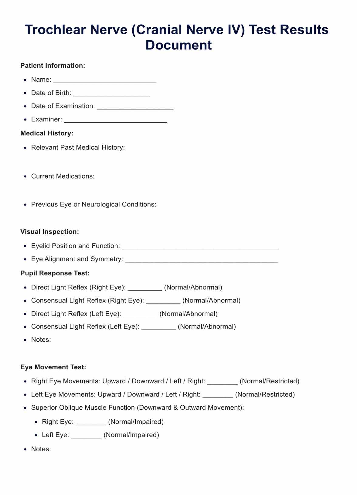
The Trochlear Nerve Test Example
What does this test measure?
The trochlear nerve is closely associated with other cranial nerves involved in eye movement, notably the oculomotor nerve (cranial nerve III) and the abducens nerve (cranial nerve VI). These nerves and the trochlear nerve facilitate the movements of the other extraocular muscles, like the lateral rectus muscle. Disruption in the synergy of these nerves can lead to complex ophthalmological presentations.
Testing the trochlear nerve typically involves examining the eye's movements, particularly looking for the ability to move the eye downward and outward. The head tilt test is a significant diagnostic maneuver in identifying trochlear nerve palsy.
In the context of the broader cranial nerve examination, the trochlear nerve is part of an intricate network that includes nerves like the trigeminal nerve (cranial nerve V), which governs facial sensation and motor function of masseter muscles, and the facial nerve (cranial nerve VII), involved in facial expressions. Other cranial nerves, such as the optic nerve (cranial nerve II), vagus nerve (cranial nerve X), and vestibulocochlear nerve (cranial nerve VIII), serve different functions ranging from sensory to motor and autonomic roles.
How does this trochlear nerve exam work?
Step 1: Preparation
Ensure all necessary equipment is available, such as a penlight, an occluder (or hand), and a target object (like a pen or finger). Arrange for adequate lighting and a comfortable space for both the patient and the examiner.
Explain the procedure to the patient, ensuring they understand and consent to the exam. Make sure the patient is seated comfortably.
Step 2: Administration of the test
Observe the patient's eyes for any obvious alignment issues or abnormal movements. Ask the patient to follow a target object with their eyes. Move the object in different directions, including the action plane of the superior oblique muscle.
Then, instruct the patient to tilt their head to both sides while focusing on a stationary object, observing for misalignment or other abnormalities. Perform the cover test on each eye individually to check for misalignment and corrective movements.
Evaluate the speed, accuracy, and smoothness of the patient's eye movements as they shift their gaze between two targets and follow a moving target.
Step 3: Post-examination
Record all findings accurately in the patient's medical record. Analyze the test results to determine if there are any indications of trochlear nerve dysfunction or other neurological issues.
Step 4: Follow-up actions
If abnormalities are detected, refer the patient to a neurologist or ophthalmologist for further evaluation. Recommend additional tests if needed, such as imaging studies (like an MRI) to investigate the underlying cause of any abnormalities.
Step 5: Continued care
Schedule follow-up appointments to monitor the patient's condition and response to any treatment or intervention. Modify the treatment plan based on the patient's progress and any new findings.
Interpreting the results
Interpreting the results of a trochlear nerve test involves a comprehensive understanding of the trochlear nerve's (cranial nerve IV) function and its relationship with other cranial nerves and anatomical structures. When analyzing the test results, fundamental aspects and keywords related to the trochlear nerve and its functioning should be considered:
- Eye movement test and superior oblique muscle function: The trochlear nerve innervates the superior oblique muscle. This muscle controls the downward and outward movement of the eye. Smooth and full-range eye movements indicate normal function in these directions. Impaired function, often seen in trochlear nerve palsy, may present as limited or strained movement, indicating a problem with the nerve or muscle.
- Head tilt test: A positive head tilt test, where tilting the head to one side worsens double vision (diplopia), can suggest trochlear nerve palsy. The affected eye may not rotate properly due to impaired superior oblique muscle function.
- Double vision (diplopia): Trochlear nerve dysfunction can lead to vertical diplopia, where the patient sees two images, one above the other. This symptom is crucial in diagnosing fourth nerve palsy.
- Other extraocular muscles and cranial nerves: The trochlear nerve works with the oculomotor (cranial nerve III) and abducens (cranial nerve VI) nerves. These nerves collectively control all the extraocular muscles, including the lateral rectus and the muscles forming the common tendinous ring. Dysfunction in these nerves can lead to complex eye movement disorders.
- Visual inspection and pupil response test: While primarily focused on motor function, examining the appearance of the eyes and pupils can provide additional context. Any abnormalities in eyelid position or pupil response, though not directly related to the trochlear nerve, can indicate broader neurological or ophthalmological issues.
- Fundoscopy: Although not directly assessing the trochlear nerve, fundoscopy can give insights into the overall ocular health, which can be pertinent in cases of trauma or systemic diseases affecting the cranial nerves.
- Patient history and context: Factors like head trauma, congenital anomalies, or raised intracranial pressure can affect the trochlear nerve. A thorough patient history can provide valuable context in interpreting test results.
- Differential diagnosis: Since symptoms like diplopia can arise from various conditions, it’s important to consider differential diagnoses. This may include assessing other cranial nerves and vestibular function and checking for signs of facial weakness, sensorineural hearing loss, or facial sensation changes.
In summary, interpreting trochlear nerve test results requires an integrated approach, considering the function of the nerve itself, its role in eye movement, and its interaction with other cranial nerves and muscles. Abnormalities in the test can point towards trochlear nerve palsy or other neurological conditions, necessitating further investigation or referral to a specialist.
Next steps
After conducting a trochlear nerve test, the next steps depend on the findings and overall clinical context. Integrating relevant keywords and concepts can provide a clearer understanding of these steps:
Normal results
If the trochlear nerve function appears normal, with no signs of double vision (diplopia), full range of motion in the superior oblique muscle, and a negative head tilt test, routine follow-up may be recommended.
In cases where there's a high suspicion of an issue despite normal test results, especially in the context of head trauma or other neurological symptoms, a cautious approach with a follow-up examination might be prudent.
Abnormal results indicating trochlear nerve palsy or dysfunction
- Further diagnostic testing: If the test suggests trochlear nerve palsy, additional tests like MRI or CT scans, especially focusing on the midbrain region, the course of cranial nerve IV, and the superior orbital fissure, might be necessary to identify underlying causes like trauma, inflammation, or tumors.
- Ophthalmologic and neurological consultations: Referral to specialists for a comprehensive evaluation, including assessment of other cranial nerves like the oculomotor (III), trigeminal (V), and abducens (VI) nerves, is crucial. These evaluations can help assess the overall impact on extraocular muscles and eye function.
- Management of symptoms: This may involve correcting double vision with prisms in glasses or patching one eye temporarily to alleviate discomfort.
Treatment of underlying causes
If an underlying cause is identified, such as a tumor in the cavernous sinus or raised intracranial pressure, appropriate treatment of the primary condition is necessary.
In congenital trochlear nerve palsy cases, observation and regular follow-up might be recommended, with surgical intervention considered in specific cases.
Rehabilitation and physical therapy
Physical therapy focusing on vestibular rehabilitation can be helpful for patients with persistent symptoms. Eye exercises might be recommended to strengthen the coordination of extraocular muscles.
Regular monitoring:
Ongoing monitoring is essential, especially in cases of traumatic injury or other neurological conditions, to assess any changes in the function of cranial nerves III, IV, and VI and to manage any emerging complications.
You can use this medical record template to improve your practice and client experiences.
Commonly asked questions
The trochlear nerve test assesses the function of cranial nerve IV (CN IV), which innervates the superior oblique muscle of the eye. This test is crucial because the trochlear nerve, the only cranial nerve that emerges from the dorsal aspect of the brainstem (near the inferior colliculus), has the longest intracranial course. It plays a crucial role in controlling the eye's movement, particularly its ability to move downward and outward. The test helps diagnose conditions like trochlear nerve palsy and can provide insights into the health of other cranial nerves and the superior orbital fissure region.
The trochlear nerve test is part of a broader examination of cranial nerves, especially those involved in eye movement, like the oculomotor (CN III) and abducens (CN VI) nerves. These nerves collectively control all extraocular muscles, ensuring coordinated eye movement. Abnormalities in the trochlear nerve test could indicate problems that might also affect the function of these other nerves and structures, like the cavernous sinus and superior orbital fissure.
Symptoms that might necessitate a trochlear nerve test include double vision (mainly vertical diplopia), difficulty moving the eye downward and outward, and a head tilt. These symptoms can arise from issues in the trochlear nucleus, the nerve's pathway, or its target muscle, the superior oblique. Conditions like acute vestibular syndrome, trauma affecting the middle cranial fossa, or issues in the lateral wall of the cavernous sinus can also prompt this test.