Sciatic Nerve Anatomy Diagram

Download our free Sciatic Nerve Anatomy Diagram PDF to understand the sciatic nerve's pathway, key landmarks, and relationships with surrounding structures.

By Olivia Sayson on Jun 21, 2024.

tick

Fact Checked by Ericka Pingol.

Use Template
Sciatic Nerve Anatomy Diagram PDF Example
toolsShare uiAI icon

What is the sciatic nerve?

The sciatic nerve is the largest and longest nerve in the human body, originating from the spinal nerves of the lower back and extending down the back of the posterior thigh of each leg. Understanding the anatomy of the sciatic nerve, including its pathway through the greater sciatic foramen and its relationships with surrounding structures like the piriformis muscle and the sacral plexus. This knowledge is essential for diagnosing and treating conditions such as sciatic nerve pain, sciatic nerve entrapment, and piriformis syndrome.

The sciatic nerve passes beneath the piriformis muscle and can be involved in piriformis syndrome if it becomes compressed or irritated. As it travels down the leg, the sciatic nerve splits into the tibial nerve and the common peroneal nerve near the popliteal fossa.

Printable Sciatic Nerve Anatomy Diagram

Download this Sciatic Nerve Anatomy Diagram to enhance your understanding and treatment of sciatic nerve conditions.

How does our Sciatic Nerve Anatomy Diagram work?

Here are five steps to effectively use the template:

Identify key landmarks

Start by familiarizing yourself with the major anatomical landmarks such as the lumbar spine (L4, L5), sacrum (S1, S2, S3), iliac bone, greater sciatic foramen, and ischial tuberosity.

Trace the sciatic nerve pathway

Follow the sciatic nerve as it emerges from the lumbosacral plexus and passes through the greater sciatic foramen.

Examine surrounding structures

Pay attention to the muscles, bones, and joints surrounding nerve roots of the sciatic nerve, such as the piriformis muscle, which can be involved in conditions like piriformis syndrome.

Identify potential compression sites

Use the diagram to locate common sites where the sciatic nerve may become compressed or entrapped, such as beneath the piriformis muscle or near the ischial tuberosity.

Apply clinical knowledge

Incorporate this anatomical knowledge into clinical practice for diagnosing, treating, and managing lower limb neuropathies, ensuring a comprehensive approach to patient care.

Sciatic Nerve Anatomy Diagram example (sample)

We have created a Sciatic Nerve Anatomy Diagram PDF example to help you understand how this free template works. This sample illustrates the anatomical pathway of the sciatic nerve. You can view the sample here or download it as a PDF for reference.

Download our free Sciatic Nerve Anatomy Diagram example here

Sciatic Nerve Anatomy Diagram example (sample)

A better solution: Carepatron

Are you ready for the best practice management software to make your life easier? Carepatron offers a user-friendly interface, powerful analytics, secure connectivity, and automated workflows to streamline your operations, save time and money, and stay connected with patients. Our software simplifies tracking patient information, storing documents, and managing communications between providers, staff, and patients.

Ready to take your practice to the next level? Explore the benefits of Carepatron's clinical documentation doftware and enhance your patient care today.

practice management software
How can the Sciatic Nerve Anatomy Diagram help in clinical practice?

Commonly asked questions

How can the Sciatic Nerve Anatomy Diagram help in clinical practice?

It helps healthcare professionals accurately identify key anatomical landmarks, trace the leg and foot sciatic nerve pathway.

What are the key landmarks highlighted in the diagram?

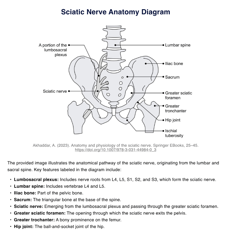
Key landmarks include the lumbar spine (L4, L5), sacrum (S1, S2, S3), iliac bone, greater sciatic foramen, hip joint, and ischial tuberosity.

How does the diagram aid in diagnosing sciatic nerve injury or entrapment?

By showing potential compression sites, such as beneath the piriformis muscle, the diagram helps in identifying areas where the sciatic nerve may become entrapped.

Join 10,000+ teams using Carepatron to be more productive

One app for all your healthcare work