Standard diagnostic tests for macular degeneration include a comprehensive eye exam, visual acuity test, dilated eye exam, and imaging tests like optical coherence tomography (OCT) and fluorescein angiography.
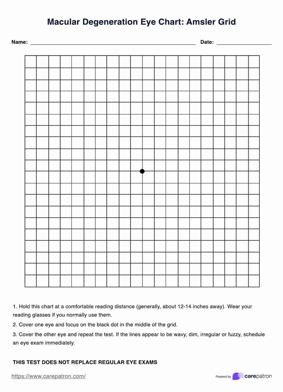
Macular Degeneration Eye Chart
Discover essential macular degeneration tests: From early detection to managing symptoms, your comprehensive guide for optimal eye health and care.

Use Template
Macular Degeneration Eye Chart Template
Commonly asked questions
Individuals aged 50 and older or those with risk factors should undergo regular eye exams, typically every two years. However, individuals with a family history, smokers, or existing eye conditions may require more frequent testing, as an eye care professional advises.
Yes, macular degeneration can often be detected in its early stages through routine eye exams. Tests like OCT can reveal signs such as drusen formation, enabling timely intervention, early diagnosis and management to slow down the condition's progression. Regular eye check-ups are crucial for early detection.


EHR and practice management software
Get started for free
*No credit card required
Free
$0/usd
Unlimited clients
Telehealth
1GB of storage
Client portal text
Automated billing and online payments