What is an Echocardiogram Stress Test?

An echocardiogram stress test, often called a "stress echo," is a non-invasive diagnostic tool that merges two tests: an echocardiogram and a stress test. This combined approach provides a dynamic assessment of the heart's functions, revealing issues that might not be evident when the body is at rest. 

During the test, an echocardiogram a heart ultrasound captures detailed images of the heart's structure and motion. When paired with a stress test that typically involves exercise on a treadmill or stationary bike, doctors can evaluate the heart's response to the added workload.

The stress echo is particularly effective in diagnosing various forms of heart disease, including coronary artery disease (CAD), the leading cause of death globally. It is also instrumental in determining the severity of heart conditions, checking the effectiveness of treatments, and predicting outcomes of diseases. During the procedure, the echocardiogram records images of the heart's chambers, valves, and pumping action before and immediately after physical exertion. At the same time, the patient's blood pressure and electrical activity are continuously monitored.

This Echocardiogram Stress Test is particularly valuable as it can unveil myocardial ischemia a condition where the heart muscle doesn't get enough oxygen-rich blood due to blocked arteries. Detecting ischemia during exercise indicates that it could also occur during everyday activities, signifying a higher risk of a heart attack. Hence, the echocardiogram stress test is a vital resource for clinicians in the diagnosis, management, and prognosis of heart diseases.

How does it work?

Step 1: Pre-Test Preparation

Patients are advised to wear comfortable clothing and shoes suitable for exercise. Electrodes are placed on the chest to monitor the heart's activity.

Step 2: Baseline Echocardiogram

An initial echocardiogram is performed to capture images of the heart at rest. The patient lies on their side while a technician uses a transducer to take pictures of the heart.

Step 3: Stress Induction

The patient then engages in physical exercise, typically on a treadmill or stationary bike, to increase the heart rate. For those unable to exercise, medication may be administered to mimic the effects of exercise on the heart.

Step 4: Exercise Echocardiogram

At peak exercise, the patient quickly returns to the table for another set of echocardiograms to capture the heart's activity under stress.

Step 5: Monitoring and Recovery

The patient's heart rate and blood pressure are monitored until they return to baseline levels.

Step 6: Analysis and Results

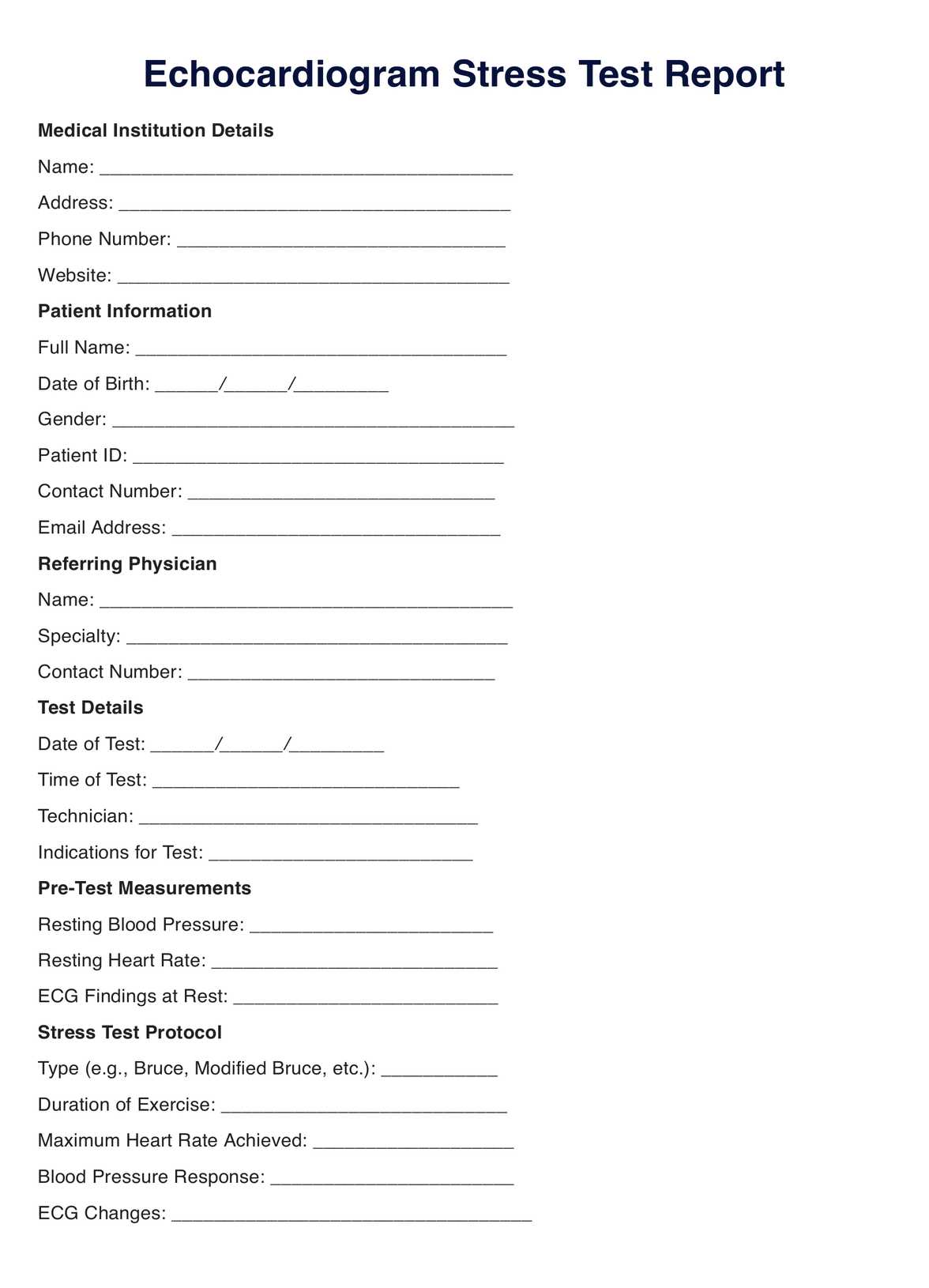
A cardiologist reviews the before and after images to assess the heart's response to stress. The results from our Printable Echocardiogram Stress Test can help diagnose various heart conditions, such as coronary artery disease.

When would you use this test?

An echocardiogram stress test is a valuable diagnostic tool healthcare practitioners utilize to assess cardiac function under physical stress. Indicated primarily for patients presenting symptoms suggestive of coronary artery disease, such as angina or dyspnea, it helps to ascertain the presence of significant heart conditions that might not be evident at rest.

Beyond symptomatic inquiry, this test is pivotal for evaluating a patient's exercise tolerance, particularly in those with known heart disorders or following cardiac procedures. It measures the heart's ability to respond to external stress, a key determinant in formulating a safe exercise regimen or gauging recovery in cardiac rehabilitation.

The echocardiogram stress test is also instrumental in monitoring treatment efficacy for various cardiac conditions. It can reveal improvements or progressions in cardiac function, guiding adjustments in therapeutic interventions, whether medical, surgical, or lifestyle modifications.

Moreover, this test is an investigative measure for the structural and functional integrity of heart valves and chambers. It identifies abnormal wall motion, valvular diseases, or heart chamber enlargement, ensuring comprehensive cardiac assessment.

Cardiologists may opt for this test in pre-operative evaluations to ensure a patient's heart can withstand surgical stress. Furthermore, in those with congestive heart failure, the echocardiogram stress test assists in carefully tailoring treatment plans and predicting outcomes.

The echocardiogram stress test is an indispensable resource for clinicians, aiding in the diagnostic and prognostic stratification of patients with cardiac concerns. It is appropriate for the clinical investigation of chest pain etiology, functional capacity assessment, treatment efficacy analysis, and detailed examination of cardiac structures and performance under duress. Its adoption in practice underscores a commitment to nuanced and informed cardiac care.

EHR software positive feedback

What do the results mean?

Doctors use an echocardiogram stress test to check how the heart works during physical activity. It's beneficial for finding heart problems that only show up when the heart works hard. This test is often for people who have signs like chest pain or trouble breathing that may mean heart disease.

It's also used to see how well people with heart conditions can handle exercise. Doctors can tell how well the heart responds to stress, which is important for planning safe workouts or seeing how well someone does after a heart procedure.

The test helps doctors track how well treatments for heart issues are working. It can show if heart function is getting better or worse, which can lead to changes in medication, surgery, or lifestyle.

This test also looks closely at the heart's structure and how well it pumps. It can spot problems with heart walls, valves, or chambers.

Before surgery, doctors might use this test to make sure a patient's heart can handle the stress of an operation. For people with heart failure, it helps doctors make personalized treatment plans and predict how the patient might do.

Overall, our Free Echocardiogram Stress Test is key for doctors to figure out the cause of chest pain, how much exercise a patient can do, how well treatments work, and to check the heart's structure and function when under stress. It plays a big part in providing detailed and careful heart care.

Research & Evidence

The echocardiogram stress test, an integration of echocardiographic imaging and physiological stress, has a well-established pedigree in cardiology, dating back to the late 20th century. Grounded in a robust body of evidence, its effectiveness and safety in diagnosing and assessing cardiac conditions are well-documented.

Historically, the stress echocardiogram evolved as a non-invasive method to detect ischemia by comparing the myocardial function at rest and under stress, usually induced by exercise or pharmacological agents. The technique gained prominence following research that confirmed its diagnostic accuracy, especially in uncovering coronary artery disease (CAD) the leading cause of morbidity and mortality globally.

Studies have consistently shown that stress echocardiography has a high sensitivity and specificity for diagnosing CAD. A landmark paper by Picano et al. (1992) demonstrated its utility in identifying myocardial ischemia and viability. Moreover, research has underscored its predictive value, with outcomes of stress echocardiograms correlating with future cardiac events, such as myocardial infarction or the need for revascularization.

Evidence also suggests that stress echocardiography is comparable to other testing modalities, like nuclear imaging, but with the added benefits of lower cost and absence of radiation exposure. Its application has expanded beyond CAD, aiding in the management of valvular heart disease, hypertrophic cardiomyopathy, and the evaluation of heart failure and pulmonary hypertension.

Recent advances, including contrast agents and three-dimensional imaging, have enhanced the test's diagnostic power. The echocardiogram stress-test continues to be a subject of ongoing research, with studies examining its utility in various clinical scenarios and its integration with other emerging diagnostic technologies.

The echocardiogram stress test is a cornerstone in the non-invasive assessment of cardiac function, buoyed by decades of evidence and continual innovation. It remains a critical tool in the cardiologist's arsenal, guiding patient management through detailed cardiac evaluation under stress conditions.

References

  1. Armstrong, W. F., & Ryan, T. (2010). Stress Echocardiography: Principles and Practice. In Feigenbaum's Echocardiography (7th ed., pp. 246-271). Lippincott Williams & Wilkins.
  2. Picano, E., Mathias, W., Pingitore, A., Bigi, R., & Previtali, M. (1992). Safety and tolerability of dobutamine-atropine stress echocardiography: a prospective, multicentre study. The Lancet, 340(8821), 722-724.
  3. Roger, V. L., Go, A. S., Lloyd-Jones, D. M., et al. (2011). Heart disease and stroke statistics, 2012 update: a report from the American Heart Association. Circulation, 125(1), e2-e220.
Who typically requests an Echocardiogram Stress Test?

Commonly asked questions

Who typically requests an Echocardiogram Stress Test?

Cardiologists or primary care physicians may request this test for patients showing symptoms of heart disease.
When are Echocardiogram Stress Tests used?

Echocardiogram Stress Tests are used when there is a need to evaluate the heart's function during physical stress.

How are Echocardiogram Stress Tests used?

Echocardiogram Stress Tests diagnose coronary artery disease, assess treatment effectiveness, and evaluate exercise tolerance.

How long does an Echocardiogram Stress Test take?

The entire procedure typically takes about an hour, including preparation, stress induction, and recovery.

Join 10,000+ teams using Carepatron to be more productive

One app for all your healthcare work