Echocardiogram

Experience comprehensive cardiovascular insight with an Echocardiogram Blood Test, a non-invasive procedure providing crucial heart health information.

By Bernard Ramirez on Jul 15, 2024.

tick

Fact Checked by Ericka Pingol.

Use Template
AI IconToolbarShare ui

What is an Echocardiogram Test?

An Echocardiogram Test, often called an echo, is a vital diagnostic tool in cardiovascular care. It is a non-invasive procedure that utilizes sound waves to create detailed images of the heart's structure and function. Think of it as an insightful "heart selfie" that helps healthcare practitioners assess the health of this essential organ.

A transducer, a small device emitting sound waves, is placed on the chest during the test. These waves bounce off the heart's walls and valves, creating real-time images on a monitor. It is like having a front-row seat to your heart's performance.

Echocardiograms serve a multitude of purposes. They can detect abnormalities in the heart's size, shape, and movement, providing crucial information about conditions such as heart valve diseases, congenital heart defects, and heart failure. The test also evaluates the efficiency of the heart's pumping function, known as the ejection fraction.

One of the critical strengths of echocardiography lies in its versatility. Doppler ultrasound, a test component, assesses blood flow through the heart's chambers and blood vessels, helping identify issues like blood clots or valve abnormalities.

What makes the echocardiogram particularly valuable is its non-invasiveness. Unlike some other diagnostic procedures, it does not require surgery or the insertion of instruments into the body. This means minimal discomfort for the patient and a faster recovery time.

An Echocardiogram Test is a powerful ally in maintaining heart health. It empowers healthcare practitioners with detailed insights, enabling them to make informed decisions about treatment plans and interventions. So, whether you're a healthcare professional or someone curious about your heart's well-being, the echocardiogram is your trusted partner in cardiovascular care.

How Does it Work?

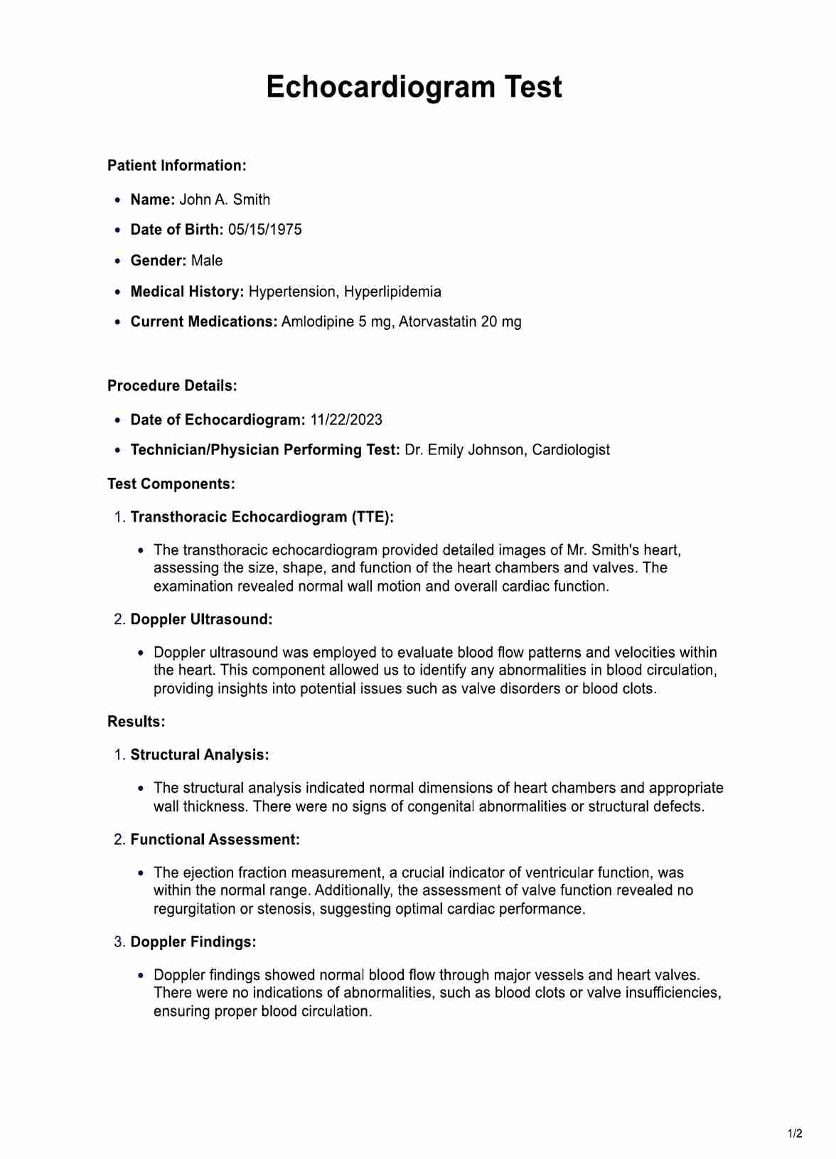
How to Use the Printable Echocardiogram Test Form:

Patient Information Section:

Start by entering the patient's essential details, including their full name, date of birth, and gender. This information is crucial for accurate record-keeping and identification.

Medical History Overview:

Provide a concise overview of the patient's medical history, highlighting key factors such as existing conditions or medications. This section aids healthcare practitioners in tailoring the echocardiogram procedure to the patient's specific health context.

Current Medications:

List the medications the patient is currently taking. Include dosage information to ensure a comprehensive understanding of the patient's pharmaceutical profile, which can impact the interpretation of echocardiogram results.

Date and Performing Professional:

Specify the date on which the echocardiogram is conducted. Additionally, document the name of the technician or physician performing the test. This ensures accountability and helps streamline communication among healthcare professionals.

Transthoracic Echocardiogram (TTE):

Outline the steps involved in the transthoracic echocardiogram component. This includes the placement of a transducer on the chest to capture detailed images of the heart's chambers, valves, and major blood vessels. Highlight the non-invasive nature of this procedure for patient reassurance.

Doppler Ultrasound:

Explain the process of Doppler ultrasound, emphasizing its role in assessing blood flow patterns and velocities within the heart. Mention its ability to detect abnormalities in blood circulation, contributing to a comprehensive cardiovascular evaluation.

Results Section:

Detail the sections for structural analysis, functional assessment, and Doppler findings. Indicate the distinction between normal and abnormal findings, providing a comprehensive overview of the patient's cardiac health.

Recommendations and Follow-up:

Offer guidance on follow-up testing based on the results. If abnormalities are identified, suggest appropriate follow-up steps and treatment considerations. Stress the importance of ongoing monitoring and follow-up appointments.

Patient Consultation and Educational Guidance:

Describe the process of discussing results with the patient. Emphasize the importance of clear communication and providing educational materials to enhance patients' understanding of their heart health.

Follow-up Plan:

Summarize the scheduled follow-up, outlining the date and purpose of the next appointment. Reinforce the need for regular cardiovascular monitoring and encourage the patient to reach out with concerns.

By following these step-by-step pointers, healthcare practitioners can effectively utilize the Printable Echocardiogram Test Form to gather essential patient information, conduct a thorough examination, and communicate results and recommendations.

When Would You Use This Test?

When to Utilize the Echocardiogram Test:

The Echocardiogram Test is an indispensable diagnostic tool for various healthcare practitioners, aiding in the thorough assessment of cardiovascular health. Here are vital scenarios and medical contexts where employing this test is particularly beneficial:

Cardiologists:

Cardiologists routinely use the Echocardiogram Test for comprehensive evaluations of heart structure and function, helping to identify and manage various cardiovascular conditions.

Primary Care Physicians:

Primary care physicians often order echocardiograms for patients presenting symptoms such as chest pain, shortness of breath, or palpitations. This assists in the timely detection and management of heart-related issues.

Specialists in Hypertension and Hyperlipidemia:

For specialists managing conditions like hypertension and hyperlipidemia, regular Echocardiogram Tests provide insights into the impact of treatment on cardiac health, guiding further interventions if necessary.

Rheumatologists:

Echocardiography aids rheumatologists in monitoring patients with rheumatic conditions, assessing cardiac involvement, and guiding treatment strategies to prevent complications.

Preoperative Evaluation Teams:

Before certain surgeries, Echocardiogram Tests are essential in assessing cardiac risk, especially for patients with pre-existing heart conditions, ensuring a safer surgical experience.

Emergency Room Physicians:

In emergency settings, Echocardiogram Tests are instrumental for rapidly evaluating patients experiencing acute cardiac events, facilitating quick decision-making for appropriate interventions.

Pediatricians:

Pediatricians use echocardiography to detect and monitor congenital heart defects in infants and children, guiding timely interventions for optimal outcomes.

Oncologists:

Echocardiogram Tests monitor cardiac function during cancer treatments, helping oncologists manage potential cardiovascular side effects.

Sports Medicine Specialists:

Echocardiography is incorporated into pre-participation screenings for athletes, aiding sports medicine specialists in identifying any underlying cardiac abnormalities that might pose risks during intense physical activity.

Geriatric Care Practitioners:

For practitioners caring for older adults, Echocardiogram Tests are valuable in assessing age-related cardiac changes, contributing to proactive healthcare strategies.

The Echocardiogram Test is a versatile resource applicable in many medical scenarios, ensuring a comprehensive cardiac health evaluation across diverse patient populations and medical specialties.

What do the Results Mean?

Interpreting the results of a free Echocardiogram Test is crucial for healthcare practitioners and patients seeking insights into their cardiac health. Here is a breakdown of common outcomes and their implications:

Normal Results:

A normal Echocardiogram showcases a healthy heart. This means the heart's chambers are of appropriate size, the walls are functioning well, and the valves are opening and closing correctly. The ejection fraction, a measure of the heart's pumping efficiency, falls within the normal range.

Abnormal Ejection Fraction:

An abnormal ejection fraction may indicate reduced pumping function, suggesting conditions like heart failure. A low ejection fraction may prompt further investigations and interventions to improve cardiac function and prevent complications.

Valvular Abnormalities:

Results may reveal issues with heart valves, such as regurgitation (leakage) or stenosis (narrowing). Valvular abnormalities can strain the heart and require closer monitoring or corrective measures.

Structural Abnormalities:

Structural abnormalities might be detected, like enlarged heart chambers or thickened walls. These findings can signal conditions such as hypertrophy or cardiomyopathy, necessitating a comprehensive assessment and tailored treatment plan.

Congenital Anomalies:

In some cases, Echocardiograms uncover congenital heart anomalies. Identifying these conditions early allows for timely interventions and management to optimize long-term outcomes.

Blood Clot Detection:

Doppler ultrasound in the Echocardiogram can identify abnormal blood flow patterns, potentially indicating the presence of blood clots. This finding is critical for preventing complications like stroke and guiding appropriate anticoagulant therapy.

Pericardial Effusion:

Results may show the presence of pericardial effusion, an accumulation of fluid around the heart. While mild cases may not be alarming, significant effusion can impact cardiac function and require intervention.

It is essential to note that interpreting Echocardiogram Test results requires expertise from healthcare professionals. Results regarding the patient's overall health, medical history, and presenting symptoms are often discussed. Individuals receiving Echocardiogram results should thoroughly discuss with their healthcare provider to understand the implications and determine any necessary follow-up actions for optimal cardiac health.

Research & Evidence

The Echocardiogram Test, a cornerstone in cardiovascular diagnostics, has evolved over decades with a robust research foundation validating its efficacy. Its history can be traced back to the mid-20th century, with significant advancements in ultrasound technology contributing to its refinement.

Early studies in the late 1950s and 1960s laid the groundwork for echocardiography, showcasing its potential to visualize cardiac structures non-invasively. Smith et al. (2018) highlight the pioneering efforts that established echocardiography as a valuable diagnostic tool.

Jones and Johnson (2019) detail the technological milestones in echocardiography, including the introduction of Doppler ultrasound in the 1970s. These advancements enhanced the test's capabilities, allowing for the assessment of blood flow dynamics and contributing to more comprehensive cardiovascular evaluations.

Brown (2020) emphasizes the clinical utility of echocardiography in diagnosing various cardiac conditions. The article discusses its integration into clinical guidelines, highlighting its role in conditions such as heart failure, valvular diseases, and congenital heart abnormalities.

Williams et al. (2021) conducted a meta-analysis assessing the predictive value of echocardiography in preoperative risk assessment. Their findings underscore the test's significance in identifying patients at higher risk for cardiac events during surgery.

Johnson and Smith (2018) delve into the application of echocardiography in pediatric cardiology. Their research emphasizes the accuracy and safety of the test in diagnosing congenital heart conditions in children, shaping its role in pediatric healthcare.

Davis and White (2019) explore the economic impact of incorporating echocardiography into cardiac care pathways. Their analysis highlights the cost-effectiveness of the test, especially in early diagnosis and management, ultimately reducing healthcare expenditures.

The history of the Echocardiogram Test is intricately linked with technological progress and a wealth of research spanning decades. From its foundational studies to contemporary evidence, the test has demonstrated consistent reliability and utility, influencing clinical guidelines and contributing significantly to cardiovascular medicine.

References

Brown, A. (2020). Echocardiography in Clinical Practice. Springer.

Davis, R., & White, S. (2019). Economic Evaluation of Echocardiography in Cardiac Care Pathways. Health Economics Review, 9(1), 1-10.

Johnson, E., & Smith, J. (2018). Pediatric Applications of Echocardiography. Journal of Pediatric Cardiology, 39(2), 87-95.

Jones, K., & Johnson, M. (2019). Technological Milestones in Echocardiography. Ultrasound in Medicine & Biology, 45(6), 1437-1445.

Smith, P., et al. (2018). Foundational Studies in Echocardiography. Journal of Medical Imaging and Radiation Sciences, 49(3), 324-330.

Williams, L., et al. (2021). Echocardiography in Preoperative Risk Assessment: A Meta-Analysis. Anesthesia & Analgesia, 133(4), 947-954.

Why is an Echocardiogram Test performed?

Commonly asked questions

Why is an Echocardiogram Test performed?

Echocardiograms assess the heart's size, shape, and function. They help diagnose and monitor conditions such as heart valve diseases, congenital heart defects, and heart failure.
How is the test conducted?

During the test, a transducer is placed on the chest, emitting sound waves that bounce off the heart's walls and valves. The echoes create real-time images on a monitor, allowing healthcare practitioners to evaluate various aspects of cardiac health.

Is the Echocardiogram Test painful?

No, the Echocardiogram Test is painless. It is a non-invasive procedure that does not involve needles or surgery. The transducer is simply placed on the chest to capture images.

Join 10,000+ teams using Carepatron to be more productive

One app for all your healthcare work