Dental Radiograph Chart

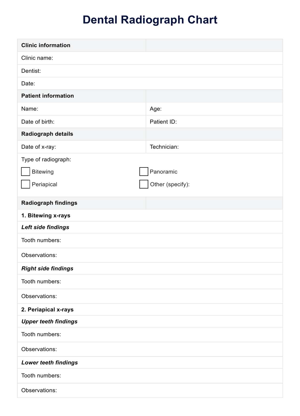
Discover how to use dental radiograph charts effectively in your practice. Download our free example and learn about the types, safety, and benefits of dental x-rays.

By Telita Montales on Jun 20, 2024.

tick

Fact Checked by RJ Gumban.

Use Template
Dental Radiograph Chart PDF Example
toolsShare uiAI icon

What is a dental radiograph?

A dental radiograph, commonly known as a dental X-ray, is a type of imaging used by dentists to view hidden dental structures, malignant or benign masses, bone loss, and cavities. These images are crucial for diagnosing and providing effective treatment planning in dental care.

What do dental radiographs contain?

Dental radiographs, or X-rays, are essential for dental diagnostics. They offer detailed views of various structures not visible during a standard examination. These images typically capture the tooth roots, jaw bones, and the bones surrounding the teeth.

By revealing hidden structures, such as the areas beneath the gums and between teeth, radiographs help dentists diagnose conditions like cavities, root infections, and bone abnormalities. They are also crucial in assessing the health of the periodontal area and the status of growing teeth and dental implants.

How much radiation exposure is there during dental X-rays

Dental X-rays involve very low levels of radiation exposure, which is considered safe for most patients. The amount of radiation emitted during dental X-rays is minimal, especially when compared to other sources of radiation encountered in daily life, such as natural background radiation from the earth and cosmic rays.

Advances in dental technology, such as digital X-rays and proper shielding with lead aprons, have further reduced the cancer risk, comparable to exposure during a short airplane flight. These safety measures ensure dental X-rays are a valuable and low-risk tool for assessing dental health issues.

Are dental x-rays safe?

Dental X-rays are considered extremely safe. They expose patients to very low radiation levels—much lower than other medical imaging procedures. Dentists use protective measures such as lead aprons and thyroid collars to ensure safety and minimize potential risks. These precautions effectively protect patients from unnecessary exposure.

Furthermore, modern dental X-ray machines limit the radiation beam to the small examined area, further reducing patient exposure. The American Dental Association and other regulatory bodies closely monitor and regulate the use of dental X-rays to ensure they are as safe as possible.

Printable Dental Radiograph Chart

Download this Dental Radiograph Chart to enhance your diagnostic accuracy and treatment planning for dental conditions.

What are the 3 types of dental images?

Understanding the different types of dental images used in radiography is crucial for accurate diagnosis and treatment planning. Here’s a closer look at the three primary types of dental X-rays commonly used in dental practices:

  1. Bitewing X-rays: Essential for evaluating the upper and lower teeth in a specific section of the mouth, bitewing X-rays capture the crowns, helping detect dental decay and changes in bone density due to gum disease. These X-rays provide a clear view of how the teeth align and interact with each other.
  2. Periapical X-rays: These images capture the entire structure of one or two teeth, from the tip of the root to the crown. They are precious for identifying issues below the gum line or in the jaw, such as impacted teeth, abscesses, cysts, or other bone abnormalities.
  3. Panoramic X-rays: Offering a broad view, panoramic X-rays capture the entire mouth in a single image. This includes all the teeth, upper and lower jaws, nasal area, sinuses, and jaw joints. They are handy for planning orthodontic treatment, assessing impacted teeth, and evaluating jaw problems.

Each type of dental radiograph uniquely enhances dental care, allowing dentists to diagnose conditions that might not be visible through a standard visual examination. Knowing when and how to use these X-rays can significantly impact the quality of care, ensuring that patients receive the most appropriate treatment for their dental needs.

How to use our Dental Radiograph Chart in treatment planning

Here's how to effectively use our printable Dental Radiograph Chart for treatment planning:

Step 1: Download our template

Start by downloading our free Dental Radiograph Chart template. This tool is designed to assist in visually assessing and planning dental treatments based on radiographic findings.

Step 2: Review radiograph images

Use the chart to review your patients' dental radiographs systematically. It will guide you in identifying and marking critical areas such as cavities, bone loss, or abnormalities in tooth roots and jawbones.

Step 3: Analyze patient information

Combine the insights gained from the radiographs with other patient information, such as medical history, oral hygiene habits, and previous dental work, to comprehensively understand the patient’s oral health status.

Step 4: Develop a treatment plan

Based on the analysis, develop a treatment plan that addresses the specific needs identified in the radiographs. The chart helps ensure that all significant findings are considered and integrated into the plan.

Step 5: Discuss with the patient

Use the chart as a visual aid to discuss the findings and proposed treatment plan with your patient. This can help explain the necessity of recommended procedures and gain the patient’s informed consent.

Step 6: Implement and monitor

After agreeing on a treatment plan, implement the necessary procedures. Use the chart during follow-up visits to monitor progress and adjust the treatment as needed.

By following these steps, you can maximize the utility of the Dental Radiograph Chart in your practice, enhancing the planning and execution of dental treatments.

Dental Radiograph Chart example (sample)

Our Dental Radiograph Chart PDF example is an invaluable resource for new and experienced dental professionals. This sample chart illustrates the common views used in dental radiography, such as bitewing, periapical, and panoramic images, linking each type of X-ray to specific dental health assessments.

The chart enhances diagnostic accuracy by demonstrating how each view should appear and what it typically reveals. It also effectively communicates radiographic findings with patients. It also shows the relationship between different dental structures captured in X-rays, helping practitioners identify and diagnose various conditions, ranging from cavities and gum disease to impacted teeth and jaw abnormalities.

This visual tool is designed to improve the understanding of dental X-rays and ensure they are used optimally in patient care. Download our free example to see how this chart can streamline your radiographic interpretations and enhance your treatment planning.

Download this free Dental Radiograph Chart example here:

Dental Radiograph Chart example (sample)

Benefits of dental radiographic examinations

Dental radiographic examinations are invaluable for many benefits, such as:

Early detection of hidden dental issues

Dental radiographic examinations are crucial in detecting conditions not visible during a standard visual examination. This includes the identification of dental caries (tooth decay) in their initial stages, subclinical gum disease, and subtle changes in bone density that could indicate the onset of periodontal problems.

Management of oral health issues

Radiographs are indispensable for diagnosing and managing a range of oral health issues. They are instrumental in planning treatments for gum disease, assessing the extent of impacted teeth, and identifying jawbone abnormalities such as cysts or tumors. These examinations provide a clear picture of the entire tooth and the surrounding bone structure, allowing for precise treatment planning.

Guidance for complex dental procedures

Radiographic examinations offer essential insights into more complex dental procedures, such as orthodontic treatments, dental implants, and root canals. They help map out the placement of dental implants, evaluate the alignment of teeth and jaws for orthodontics, and navigate through root canal procedures by revealing the shape and number of root canals.

Monitoring the progress of dental treatments

Regular dental X-rays are also vital in monitoring the progress of ongoing treatments. They can track the evolution of dental and periodontal diseases, verify the success of dental restorations, and ensure that no new issues have developed. This ongoing assessment helps make timely adjustments to the treatment plan, ensuring optimal outcomes for the patient.

These benefits highlight why dental radiographic examinations are a cornerstone of modern dental practice. They significantly enhance the ability to diagnose, plan, and monitor dental treatments effectively.

Why use Carepatron as your dental practice software?

Carepatron's patient portal software is designed to streamline dental practice operations, integrating essential tools for managing dental radiographs effectively. Our telehealth platform enhances diagnostic processes by providing quick access to radiographic data, improving the accuracy and efficiency of treatment planning.

Furthermore, Carepatron ensures that all radiographic images and patient data are stored securely, complying with privacy regulations to protect patient information. This comprehensive approach simplifies daily workflows and supports dental professionals in delivering superior patient care. Learn more about how Carepatron can enhance your practice by exploring our health coach software.

dental practice software
Are dental X-rays expensive?

Commonly asked questions

Are dental X-rays expensive?

Dental X-rays are generally affordable and are often covered by dental insurance as part of routine dental care.

Is a dental X-ray painful?

No, dental x-rays are painless. The process involves minimal discomfort, as it is non-invasive and quick.

How often should dental X-rays be taken?

The frequency of dental X-rays depends on the individual's oral health condition, age, risk for dental disease, and any signs of oral disease. Dentists typically recommend X-rays during routine dental check-ups.

Join 10,000+ teams using Carepatron to be more productive

One app for all your healthcare work