AI IconToolbarShare ui

What is a cavovarus foot deformity?

The human foot is a complex and intricate structure composed of numerous bones, joints, and over 100 muscles, tendons, and ligaments. This elaborate network of components works harmoniously to support the body's weight, facilitate movement, and maintain balance. Cavovarus foot deformity is a condition that disrupts this delicate balance and function.

Cavovarus foot deformity is characterized by an unusually high arch (cavus) and an inward tilt of the heel bone (varus), causing the foot to deviate from its normal alignment. This condition can lead to uneven weight distribution across the foot, primarily placing excess pressure on the ball and heel. As a result, individuals may experience pain, particularly on the lateral (outer) side of the foot, and instability in the ankle.

The deformity can cause challenges with walking and standing, impacting gait and overall mobility. Over time, the condition may worsen if not addressed, potentially leading to further complications such as chronic pain or repeated ankle injuries. While the underlying causes of cavovarus foot deformity vary and may include congenital factors, neurological disorders, or traumatic injuries, prompt and appropriate management can help alleviate symptoms and improve the quality of life for those affected.

Characteristics of this foot deformity

Here are some signs when patients have the characteristics of cavovarus foot deformity:

  • Heel varus: Inward tilting of the heel, leading to foot instability.
  • High plantar arch: Abnormally high arch that is visible from the side.
  • Forefoot pronation: Outward tilt of the forefoot.
  • Lateral foot pain: Discomfort on the outer side of the foot.
  • Gait disturbances: Abnormal walking patterns.
  • Lateral ankle instability: Prone to ankle sprains and instability.

Risk factors and causes of this foot deformity

Here's a breakdown of the risk factors and causes of cavovarus foot deformity:

  • Congenital defect: Some individuals are born with cavovarus foot deformity due to genetic factors.
  • Neurological disorders: Conditions such as cerebral palsy and muscular dystrophy can result in muscular imbalances and foot deformities like cavovarus foot.
  • Charcot-Marie-Tooth disease: This genetic disorder affects the peripheral nerves, leading to muscle weakness and deformities such as cavovarus foot.
  • Stroke or spinal injury: Damage to the nervous system from a stroke or spinal injury can cause muscle imbalances, leading to cavovarus foot.
  • Traumatic injury: Trauma or injury to the foot, ankle, or nerves can also lead to the development of cavovarus foot deformity.

What is the Coleman Block Test?

The Coleman Block Test is a clinical assessment used to evaluate the flexibility of the hindfoot in individuals with cavovarus foot deformity. During the test, the patient stands with the outer (lateral) side of the foot resting on a 1-inch block while the inner (medial) side of the foot and first metatarsal head remain off the edge of the block, allowing them to hang freely. This positioning helps practitioners observe how the hindfoot responds when the medial forefoot is not bearing weight.

As the patient stands in this position, practitioners assess the alignment of the hindfoot to determine its flexibility. Specifically, they observe whether the heel corrects from an inward tilt (varus) to a neutral position. This assessment can provide insight into whether the hindfoot or the forefoot primarily drives the deformity.

How does our Coleman Block Test template work?

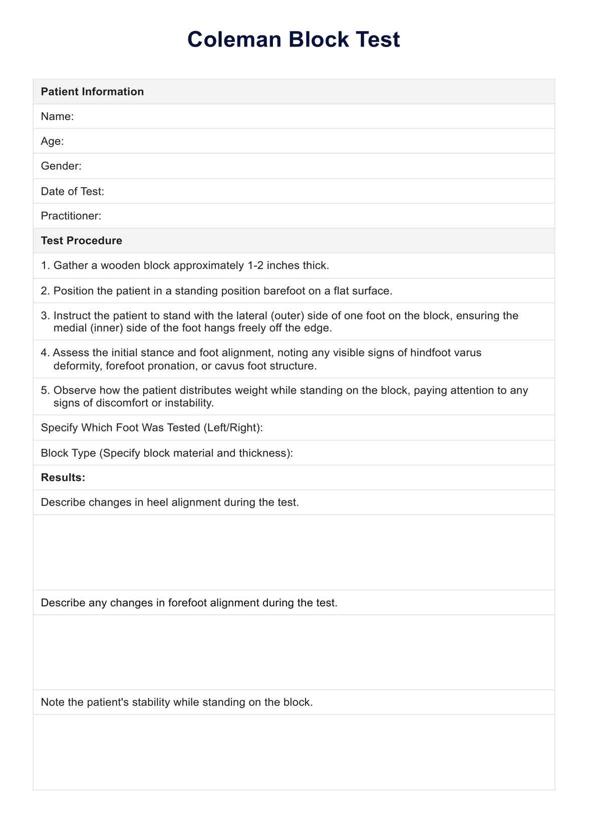
Documenting the actual physical test ensures thorough and consistent reporting. This template aids in recording the patient's initial stance, any changes in hindfoot alignment during the test, and other relevant observations that may guide diagnosis and treatment planning. Here's how our template works:

Step 1: Access the template

Begin by accessing the Coleman Block Test documentation template to ensure you are prepared to record the test results accurately. The readily available template will help guide your observations and allow for consistent and thorough documentation throughout the test.

Step 2: Patient information

Record the patient's information at the start of the test, including their name, age, gender, and the test date. Note the practitioner's name and any pertinent medical history that may influence the test results or the patient's ability to complete the test.

Step 3: Prepare for test

Gather the necessary materials, such as a 1-inch block, and position the patient with the lateral (outer) side of the foot on the block, allowing the medial (inner) side and the first metatarsal head to hang freely. Instruct the patient to stand comfortably and maintain balance during the test.

Step 4: Record observations

As the patient stands on the block, carefully observe and record their stance, foot alignment, and any heel and forefoot position changes. Note the patient's stability, reported discomfort, and other notable observations during the test.

Step 5: Record results and interpretation

Summarize the test results by noting changes in hindfoot flexibility and alignment and forefoot alignment. Interpret the results to determine whether the deformity is driven by the hindfoot or forefoot, guiding the patient's treatment plan. Based on your interpretation, document any recommendations for further assessment, treatment options, and follow-up appointments.

Benefits of having a documentation tool while conducting this test

Using a documentation tool while conducting the Coleman Block Test offers numerous benefits that enhance the assessment process for cavovarus feet:

  • Accurate recording: A structured template lets you systematically record observations such as the resting calcaneal stance position, hindfoot varus deformity, and the medial malleolus position, ensuring nothing is overlooked.
  • Clear interpretation: By documenting changes in hindfoot flexibility, forefoot pronation, and lateral forefoot alignment, you can easily interpret the results and understand the extent of cavus foot or cavovarus deformity.
  • Efficient surgical planning: Thoroughly recorded data can facilitate surgical planning if necessary, helping surgeons understand the specific areas of concern, such as hindfoot deformity and weight-bearing issues.
  • Consistent patient care: Consistency in recording findings such as the lateral aspect of the foot and how the first metatarsal head can hang freely on the block allows for standardized assessment across different patients.
  • Improved communication: Clear documentation helps practitioners effectively communicate their findings and treatment recommendations to other healthcare providers and patients.
Can cavovarus foot deformity worsen over time?

Commonly asked questions

Can cavovarus foot deformity worsen over time?

Yes, without appropriate management, the condition can worsen over time, leading to increased pain, mobility issues, and a higher risk of injuries such as ankle sprains.
Can lifestyle changes help manage cavovarus foot deformity?

Yes, wearing supportive footwear, using orthotics, and engaging in targeted exercises can help manage the symptoms and improve foot function. Physical therapy may also help strengthen the muscles around the foot and ankle.

When should I seek medical attention for cavovarus foot deformity?

You should seek medical attention if you experience persistent pain in the foot or ankle, frequent ankle sprains, difficulty walking, or any noticeable changes in gait or foot alignment.

Join 10,000+ teams using Carepatron to be more productive

One app for all your healthcare work